Medical expert of the article

New publications
Why do brown spots appear on the body and what to do?
Last reviewed: 29.06.2025

All iLive content is medically reviewed or fact checked to ensure as much factual accuracy as possible.
We have strict sourcing guidelines and only link to reputable media sites, academic research institutions and, whenever possible, medically peer reviewed studies. Note that the numbers in parentheses ([1], [2], etc.) are clickable links to these studies.
If you feel that any of our content is inaccurate, out-of-date, or otherwise questionable, please select it and press Ctrl + Enter.
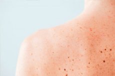
If brown spots on the body (on the fur) of a leopard, cheetah or spotted hyena in their habitat serve as camouflage for them, then on the human body such "marks" clearly have no function and in some cases indicate serious pathology...
Causes of the brown spots on the body
Brown spots on the body appear for a variety of reasons. First of all, hyperpigmentation of the skin, which dermatologists may call melanosis or melanoderma, is the result of overexposure to ultraviolet radiation. [1]
Ultraviolet (UV) sunlight activates melanocytes in the skin's basal layer - cells whose special organelles, melanosomes, produce the protective pigment melanin. Melanosomes are carried by dendrites to the upper layer of keratinocytes (which are located in the epidermis) and accumulate around their nuclei in the form of melanin caps - to mitigate UV damage to DNA. The longer and more intense the UV exposure, the more supranuclear melanin the keratinocytes accumulate: this is how pigment spots - brown spots on the body after sunbathing - are formed.
One of the acquired UV-induced skin pigmentation disorders is Riehl's melanosis, which has the appearance of numerous small or reticulated brown spots on the upper chest, neck, and face. It was previously called toxic melanoderma, but after clarification of the secondary nature of this pigmentation disorder to contact dermatitis and the identification of a delayed-type hypersensitivity reaction, a new classification has recently been adopted: acquired dermal macular (i.e., patchy) hyperpigmentation. [2]
Brown spots of various sizes and shapes on the face and body, including brown spots on the abdomen or back, can occur as so-called post-inflammatory hyperpigmentation. For example, this occurs in cases of hypersensitivity (sensitization) to solar ultraviolet light, defined as sun allergy, which may be due to the use of medications with phototoxic effects, contact with photosensitizing plants or certain substances. Symptoms of photodermatitis inflamed areas of skin - hyperemia, pustule formation, pruritis and subsequent ulceration - disappear as they heal, but brown patches remain in place of the inflammation. [3]
By the way, post-inflammatory hyperpigmentation is observed in many skin problems, including inflammatory dermatoses with fungal or viral lesions, acne, dermatitis (allergic contact and atopic). For example, shingles caused by Varicella zoster virus and red flat lichen planus, after the inflammation heals, give spots of all shades of brown color at the sites of burst blisters. Brown crusted spots on the body may be one of the consequences and complications of streptoderma.
In superficial mycoses, e.g. variegated rash, which is caused by the lipophilic yeast-like fungus Pityrosporum cibiculare of the genus Malassezia present on the skin, light, dark, pale, red, red, white, pink-brown spots appear on the body. Increased humidity, heat and sun exposure can aggravate this fungal disease. [4], [5]
This is followed by focal hyperpigmentation in various systemic diseases, which include:
- Primary or chronic adrenal insufficiency (hypocorticism, hypoadrenocorticism, or Addison's disease);
- Hypercorticism - Icenko-Cushing's syndrome;
- Caused by gene mutations neurofibromatosis (Recklinghausen's disease);
- Hyperthyroidism (thyrotoxicosis);
- Tuberous sclerosis;
- Primary biliary cirrhosis.
Quite often this form of skin pigmentation disorder is a side effect of photosensitizing drugs of various pharmacological groups.
Risk factors
In addition to UV exposure, risk factors for hyperpigmented spots on the skin include:
- Genetic predisposition;
- Dermatologic diseases, including inflammatory diseases;
- Hormonal changes;
- Thyroid disease;
- Endocrine disorders;
- Metabolic disorders;
- Autoimmune diseases;
- Drug therapy with photosensitizing drugs;
- Inappropriate skin care products and methods.
Pathogenesis
The mechanism of pigment spots formation under the influence of ultraviolet light has been described above, only it should be added that UV radiation from the sun causes lipid peroxidation in cell membranes, and the resulting free radicals stimulate melanogenesis. Also note that the mechanism has two variants: melanocytosis - an increase in melanin content with an increase in the number of functioning melanocytes in the skin, and melanosis - an increase in melanin synthesis without an increase in the number of melanocytes. Both variants may be present at the same time.
In addition, the female sex hormones estrogen and progesterone, adrenal cortex steroids, thyroid hormones, and the middle lobe of the pituitary gland alpha-melanotropin (α-MSH) are also involved in melanogenesis. This hormone is also produced and secreted by melanocytes in the skin in response to ultraviolet light, where it increases melanin synthesis.
Excess thyroid hormones activate melanocyte growth, while estrogen and progesterone can stimulate melanocyte proliferation and induce melanogenesis, increasing melanin content in the skin with subsequent hyperpigmentation.
The exact pathogenesis of postinflammatory hyperpigmentation is not yet fully understood, but it is attributed both to the cause of the inflammatory process and its chronic and/or recurrent nature, and to damage to the basal membrane of the epidermis. It is likely that hyperpigmentation is caused not only by increased melanogenesis, but also by an abnormal distribution of produced melanin, when basal keratinocytes do not retain melanocytes, and those, moving upwards, cause spontaneous pigmentation in the uppermost layer of keratinocytes.
In Addison's disease, hyperpigmentation is a chain of pathological hormonal factors: insufficient steroid production by the adrenal cortex → increased production of adrenocorticotropic hormone (ACTH) → increased biosynthesis of alpha-melanotropin → expression of MC1 skin melanocyte receptor alpha-MSH → increased melanocyte activity and additional melanin synthesis. [6]
This is also how skin manifestations in Cushing's syndrome are explained.
Symptoms
Some varieties of focal hyperpigmentation include oval-shaped flat brown patches on the body with a well-defined edge surrounded by normal-looking skin - lentigo. Simple lentigos are benign melanocytic hyperplasia with a linear distribution: the hyperplasia is confined to the layer of cells immediately above the basal membrane of the epidermis, where melanocytes are normally found.
Small brown spots on the body of the elderly are sun lentigos, also called senile lentigos, age spots, and liver spots, although they have nothing to do with liver disease: they just have a similar color (brown to dark brown) to the liver. These spots increase with age and may cluster, especially in areas that are repeatedly exposed to ultraviolet radiation (the back of the hands, top of the feet, face, shoulders and upper back).
Brown spots on the body in women
Pigment spots during pregnancy - melasma or chloasma spots (chloasma gravidarum) or the "mask of pregnancy" - are associated with increased levels of the female sex steroids estrogen and progesterone, as well as alpha-melanotropin (α-MSH). During pregnancy, α-MSH levels increase, virtually maintaining adequate levels of prolactin necessary for lactation. Blemishes of all shades of brown with irregular borders are localized on those parts of the body that are most exposed to sunlight. [7]
Melasma also appears in women who are taking oral contraceptives (birth control pills) with estrogen or are undergoing hormone replacement therapy.
Epidermal melasma results from an increase in melanin in the suprabasal layers of the epidermis, while dermal melasma results from an excess of pigment in the macrophages of the dermis.
Brown spots on the body in men
These spots in men can be:
- Brown flat moles (nevi) that are located anywhere on the skin. They are not affected by UV light and do not increase in size or darken;
- Becker's nevus, which appears in adolescence on the upper arm, anterior chest or back as a large brown spot with subsequent hair loss; [8]
- Lentigos are brownish-brown spots that get bigger over the years.
And in men with hyperhidrosis (excessive sweating), brown sweat spots may appear on the body from contact with petroleum or coal distillation products - under the influence of heat and light. This is a symptom of pigmented contact dermatitis (a non-exematous form of contact dermatitis) called Hoffmann-Habermann toxic melanoderma.
The baby has brown spots on his body
Children as young as a few months of age may have patchy papular rashes (on any part of the body) due to pigment urticaria (cutaneous mastocytosis). The red-brown, yellow-brown, and brown mole-like spots on the body are itchy; over time, the spots become larger but less itchy, and by adolescence, most of the spots disappear. Pigmentary urticaria is caused by an excessive number of mast cells in the skin - mastocytes - which, when rubbed, exposed to heat or any other irritation, produce histamine, which initiates immediate-type allergic reactions and causes itching. As it turned out, the pathogenesis of most cases of pigment urticaria is associated with a point mutation in the gene of one of the amino acids of the transmembrane protein CD117.
Small brown spots on the body can be a symptom of hereditary xeroderma pigmentosum. [9] And Recklinghausen's disease is characterized by a significant number of smooth light brown (coffee-and-milk-colored) small oval-shaped spots on the torso. As the child grows, the number and size of the spots increase. [10]
Round brown spots on the body are most commonly moles (or nevi). Read more:
Small brown spots on the body and face - freckles - are also the result of the skin being exposed to ultraviolet rays with an increase in melanin in the epidermal layer of the skin.
Large brown spots on the body may be congenital melanocytic nevi. In people with weakened immune systems, HHV-8 (human herpes virus type 8) can cause a form of cancer with the development of atypical cells around lymph nodes and blood vessels called Kaposi's sarcoma. And with this disease, skin lesions can appear: purple and red spots of varying sizes, as well as large brown spots on the body. And large "coffee-and-milk" colored spots are seen in people with tuberous sclerosis.
In most cases, protruding brown spots on the body are intradermal or convex moles, aka epidermo-dermal nevi. They can also be warty or verrucous nevi.
Hyperpigmented itchy plaques with wavy surface and brown spots without clear borders on the back (between the shoulder blades) are symptoms of primary macular cutaneous amyloidosis (deposition of fibrillar amyloid protein in the dermis).
Brown spots on the legs may be a sign of purpura pigmentosa progressiva - hemosiderosis of the skin or Schamberg's disease, as well as acroangiodermatitis associated with chronic venous hypertension. [11]
Also read:
Diagnostics of the brown spots on the body
The diagnosis involves a thorough patient examination and history - asking about all medications taken.
Instrumental diagnosis in dermatology is performed using:
- Dermatoscopy;
- With a Wood's lamp examination;
- Ultrasound of skin and subcutaneous fat;
- Siascopies.
Tests such as clinical blood tests, immunoglobulin tests (IgG, IgM, IgA), hormone levels, herpes testing, etc. Are required.
Differential diagnosis
Differential diagnosis should distinguish melasma from post-inflammatory and drug-induced hyperpigmentation, and lentigos from freckles, etc.
Treatment of the brown spots on the body
Given the wide range of causes of macular hyperpigmentation, treatment should include drugs aimed at therapy of the underlying disease. By the way, freckles and moles, as well as solar lentigo of the elderly is not a skin disease.
How to remove brown spots on the body? Detailed information in the articles:
What is the right cream for brown spots on the body, read in the publications:
- Creams for age spots
- Whitening creams for age spots
- Whitening creams for face from age spots and freckles
Physical therapy treatments include chemical peels, laser and cryotherapy.
Used phytotherapy - treatment with herbs: decoctions, infusions and extracts of such plants as chamomile (flowers), parsley (greens), dandelion (leaves), calendula (flowers), licorice (root).
In some cases, surgical treatment is also used, see more details:
Complications and consequences
If a brown spot on the body grows, experts consider it an independent risk factor for developing melanoma.
Prevention
As preventive measures, dermatologists recommend avoiding the sun at the height of the day and applying sunscreen.
Forecast
Melasma or chloasma that occurs during pregnancy is bound to go away after childbirth, although not immediately. In some patients, brown spots on the body may also disappear spontaneously over time, but this does not apply to senile lentigos, cases with endocrine problems, or syndromal conditions.