Medical expert of the article
New publications
A convex mole: what do you need to know about it?
Last reviewed: 04.07.2025

All iLive content is medically reviewed or fact checked to ensure as much factual accuracy as possible.
We have strict sourcing guidelines and only link to reputable media sites, academic research institutions and, whenever possible, medically peer reviewed studies. Note that the numbers in parentheses ([1], [2], etc.) are clickable links to these studies.
If you feel that any of our content is inaccurate, out-of-date, or otherwise questionable, please select it and press Ctrl + Enter.

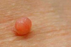
A convex mole (nevus) is a benign neoplasm on the skin. From the point of view of dermatologists, moles and birthmarks have similar medical causes. At the initial stage of development, a mole looks like a small dark spot on the skin. Later, it can remain flat or rise above the level of the skin, that is, it becomes convex. It all depends on the location of the pigment cells. If the melanocytes are located in the epidermis (the upper layer of the skin), the mole will remain flat. The mole becomes convex when the pigment cells are located in the deep layers of the skin (dermis).
Causes convex mole
A convex mole is formed due to pathological changes in the skin (proliferation of cells, resulting in the formation of a growth or thickening of the skin of a certain radius). Sometimes a birthmark may contain melanin pigments, giving it a darker shade of color. Melanin is synthesized in the presence of melanocyte cells due to exposure to ultraviolet radiation. It is worth noting the role of melanotropic hormone, produced by the pituitary gland. Therefore, more than one body system is involved in the process of pigmentation of moles.
The main reasons are the presence of certain factors – local developmental defects, hereditary predisposition, ultraviolet radiation, hormonal imbalance in the body, injuries, infections and viruses.
Local causes of developmental defects
We are talking about birthmarks of a congenital nature, which in 60% of cases are the cause of pigment spots. In this case, a convex birthmark appears as a result of a violation of the correct division of cells in the last stages of pregnancy. Basically, such a defect is hardly noticeable at the birth of a child. And only after 2-3 years does the neoplasm manifest itself during a visual examination.
Hereditary factors
At the moment, it is impossible to exclude the fact that birthmarks may appear due to hereditary skin pathologies. Tumors and birthmarks are initially coded in certain genes in the deoxyribonucleic acid (DNA) molecule. This genetic chain is transmitted to children from parents via chromosomes.
Nevi caused by exposure to ultraviolet radiation
The growth of melanocytes in the basal layer of the skin is stimulated by ultraviolet radiation. With increased insolation, the number of melanocyte cells will increase. Thus, if you want to get a dark skin tone (tan), which occurs as a result of the normal reaction of cells to sunlight, there is a risk of pathological changes in the cells of the epidermis and dermis. The appearance of such convex moles is typical for adults and is acquired.
Hormonal factor
Medical observation of patients with convex moles has shown that hormones take an active part in the process of nevi formation. According to the research results, it was noted that most often, moles of an acquired nature appear in adolescents during the maturation of the reproductive system of the body. People with serious failures in the endocrine system are also at risk. In rare cases, women are susceptible to the appearance of neoplasms during pregnancy. The main reason is the influence of hormonal changes of a pathological or physiological nature on the functions of the pituitary gland. In these cases, the prognosis of the disease is favorable, since such moles are small in size and can disappear on their own some time after their appearance.
Trauma, bacterial infections and viruses
The traumatic factor of nevi formation (mechanical damage, insect bite) is a secondary and rare cause. In this case, the main role is played by the inflammatory process in various layers of the skin. As a result of inflammation, biologically active substances are formed that stimulate cell proliferation. A similar mechanism of formation has convex moles that occur as a result of viral and bacterial infections entering the body. It should be noted that when a person is infected with the papilloma virus, the resulting convex mole differs in its nature. Therefore, from the point of view of histology and dermatology, it is classified as a papilloma, not a nevus.
The above factors of occurrence of convex moles allow to determine the risk group. This includes people predisposed to occurrence of moles. Convex moles, which are prone to develop into malignant neoplasms, are especially dangerous.
Risk factors
Who may be at risk:
- people working in industries with high ultraviolet radiation;
- people involved in the chemical or other industries in the production process of which carcinogenic substances are used;
- people who often vacation in southern (equatorial) countries;
- people suffering from chronic endocrine diseases;
- people with low immunity;
- people with diseases whose treatment requires long-term use of hormonal drugs;
- people born with a large number of nevi, as this factor can lead to the appearance of new convex moles with subsequent transformation into cancer;
- people who have relatives with histological confirmation of a diagnosis of melanoma (skin cancer);
Of all the types of moles (about 50), about 10 varieties of nevi are most common. They are divided into melanin-non-hazardous or melanin-hazardous formations. The first type includes moles without a predisposition to transform into skin cancer. Their removal is only for cosmetic purposes. The other type of nevi is dangerous because benign cells can begin to transform into malignant ones at any time.
 [ 6 ]
[ 6 ]
Symptoms convex mole
A raised mole on the face
Usually, convex moles on the face do not pose a particular danger. The only factor aimed at getting rid of a nevus is a cosmetic defect. If a mole causes a feeling of discomfort, it should be removed. Today, the procedure for removing convex moles on the face does not pose any particular problems. But you should choose the removal method with caution, taking into account the features of the facial skin.
The surgical method is not suitable for aesthetic reasons, as it may leave scars on the face. The radiosurgery method is effective only in removing small moles. Liquid nitrogen exposure (cryodestruction) is a process that does not require large financial costs, but takes a long time. The electrocoagulation method carries the risk of damaging the nevus, which can lead to malignant transformation of the mole. A safer option is the laser surgery method. That is, none of the methods for removing convex moles on the face is 100% safe. To gain complete confidence in the correctness of the choice, you need to contact a qualified surgeon for professional advice.
 [ 10 ]
Raised moles on the nose
A convex mole on the nose can be considered dangerous, as it is subject to constant risk of mechanical damage (contact with a handkerchief, rubbing with glasses, etc.). This is an unfavorable factor that acts as a starting point for the inflammatory process of the mole, transforming it into melanoma or skin cancer. Another risk factor is the adverse effects of ultraviolet rays. After all, no one uses protective equipment for the nose in everyday life.
Should a convex mole on the nose be removed? If the mole does not bother you and looks aesthetically pleasing, then it is not necessary to remove it. In cases where the mole changes color, structure and shape, you should think about getting rid of the nevus forever. The methods for removing a convex mole on the nose are the same as on the face.
A raised birthmark on a child
Recently, many young mothers have been worried about the presence of convex moles on their children. It has been proven that one child out of a hundred is born with moles, in other cases, moles appear much later (approximately at the age of 5 to 6 years). A convex mole on a child looks the same as on adults. Basically, these are nevi up to 1 cm in diameter and light brown in color. Often, such formations do not pose a danger to the child's health.
It is another matter if a convex mole begins to behave unusually - it quickly grows in size, changes color, bleeds or peels. In this situation, you need to consult a specialist. Another alarming symptom is an intensive increase in the number of moles. Today, doctors rarely insist on emergency surgery. This is explained by the peculiarities of a growing organism. Conservative treatment is more often advised. But there are situations in which (for medical reasons) it is necessary to remove a nevus urgently.
The most common method of removing a convex mole in a child is laser surgery. The operation itself is absolutely safe. Children tolerate it quite well. It is more important to pay attention to the postoperative period. It is recommended to resort to a gentle regimen, as well as to taking medications to normalize the immune system. It is necessary to limit the child's exposure to the sun, taking water procedures until the skin is completely healed. To prevent any complications, it is necessary to periodically undergo dermatological examinations. This is especially true during puberty.
Well-known facts about birthmarks
- Convex moles are most often congenital.
- Moles have the property of changing the color of skin pigmentation.
- In women, nevi are more common (both skin and mucous membranes are affected).
- Many people do not suspect that the presence of the papillomavirus, which is present in 85% of the population, is the cause of the development of convex formations similar to moles.
- Certain types of birthmarks reach quite large sizes (more than 30 cm), which reduces the quality of life of many people, causing cosmetic defects and psychological problems.
- Benign skin neoplasms are especially often transformed into malignant ones in people who have light-colored hair and eyes.
- But there is also a positive fact - many nations believe that people with a large number of birthmarks are luckier.
Forms
Types of non-melanoma-prone convex moles
Intradermal pigmented mole
Basically, this type of nevi develops in adolescence. In the initial stage, it is localized in the deep layers of the dermis, without protruding beyond its borders. The size of the mole is several millimeters. The most common location is the skin in the throat and neck area, under the chest, in the skin folds of the armpits and groin. Over time, this convex mole can slightly change in shape and color.
The prognosis is favorable. Malignant transformation (malignancy) occurs in approximately 15% of cases in the presence of additional risk factors.
Papillomatous mole
A characteristic feature is a pronounced elevation on the skin surface, differing in shape and color. Visually, it looks like a bumpy brown or pink convex mole with a granular surface. When palpated, it turns out to be soft and painless. Usually, apart from a cosmetic defect, it does not cause much concern. The location is mainly the scalp. Very rarely, it can be located on the trunk and limbs.
The prognosis is favorable. A papillomatous mole tends to gradually increase in size throughout a person's life, but cases of malignant transformation are very rare.
Sutton's nevus (halonevus)
In appearance, it is an oval or round pale convex mole. A characteristic difference is a halo of pale skin surrounding the base of the nevus. The predominant location is the skin of the limbs or body. Sometimes it can be localized on the feet, mucous membranes and face. It is necessary to take into account that when one mole of this type appears, similar ones should be looked for, since single manifestations of this type of formation are not typical.
The prognosis is favorable. The neoplasms themselves disappear without treatment after several months from their appearance. Therefore, their removal is not recommended. The transformation of halo nevus into skin cancer occurs in very rare cases. However, these moles may indicate the presence of other serious diseases that should be diagnosed in a timely manner.
Types of melanoma-prone convex moles
Blue nevus
Blue nevus (Jadassohn-Tice or blue) is considered a type of precancerous tumor, but mainly refers to the type of benign formations. The nevus received its name due to its cells that actively produce melanin. Externally, it is a dark (dark blue, dark purple) or black convex mole. This neoplasm does not have clear location statistics. The mole does not exceed 1 cm in diameter. Blue nevus is not characterized by hair growth on the surface. A more thorough examination reveals clear borders of the mole and tightness of the skin.
The prognosis is favorable. Cases when moles of this type develop into skin cancer are rare. Most often, this happens after unsuccessful removal or injury to the formation. However, people with blue nevus are recommended to undergo regular and timely preventive examination by a dermatologist.
Giant pigmented mole
This type of nevi differs from other types in that it is congenital and noticeable already in the first days of a newborn's life. External signs during examination are a large gray or brown convex mole. With the development of the body, it tends to increase significantly (from 2 to 7 cm). In some cases, it is located on large areas of the skin of the body (cheek, neck, a significant part of the body). Quite often, intense hair growth is noted on the bumpy surface of a convex mole.
The prognosis is favorable. Surgical treatment of this type of moles is prescribed to eliminate the cosmetic defect. However, cases of transformation into malignant tumors are not so rare (about 10%). The reason for this phenomenon is the large size of the mole localization area, which increases the likelihood of their injury.
Complications and consequences
Is a raised mole dangerous?
Basically, a convex mole does not pose a particular danger and does not require surgical intervention. Most people live with nevi all their lives without feeling any discomfort. Moreover, in old age, moles often disappear, transforming into pigment spots. However, some types of moles are precancerous diseases. This is the danger of nevi.
Diagnostics convex mole
- Patient interview (anamnesis collection). First of all, the family history is studied. It is clarified whether blood relatives have birthmarks and convex moles. The question about diagnosed melanoma among family members is also specified. The question about the presence of the above-mentioned external and internal risk factors in the patient's daily life is necessarily asked.
- Visual examination data. The neoplasm is assessed according to certain criteria: size and number of nevi, consistency and color, time of appearance and localization, changes that have occurred since the last medical examination.
- Dermatoscopy. It is performed using a special medical device that magnifies the image of the material being examined several dozen times. Thanks to this, the specialist can notice the smallest changes on the surface of a convex mole.

- Thermometry. Using a special device, local measurement of skin temperature is performed. During the study, the temperature of healthy skin and the surface temperature of a convex mole are compared.
- Biopsy. Used at the final stage of diagnostics, when other methods of research have already been carried out, and the diagnosis has not yet been made. An alternative to this method is cytological analysis. It is carried out by scraping off the cells of the mole. If there are secretions or ulcers on the surface of the nevus, the sample is taken by applying a glass slide to the neoplasm.
Tests
Such tests as a biochemical blood test, a general blood test and a urine test are usually not prescribed for diagnostic examination of convex moles. This is due to the fact that there are no characteristic changes in these neoplasms. In order to study the functioning of the patient's internal organs, these tests are carried out before a biopsy or before surgery to remove a convex mole. If moles appeared as a result of infections or chronic diseases, the tests are repeated. This is due to the need for proper treatment, since in this case the nevus is a symptom and does not require emergency treatment.
Who to contact?
Treatment convex mole
Treatment of convex moles begins after diagnostics, including a biopsy of suspicious tissue. Medication is ineffective in cases of already formed formations, so it is practically not used. Drug treatment is prescribed in cases where nevi have formed against the background of other diseases.
Treatment methods for raised moles:
- surgical removal of nevi;
- treatment with folk remedies;
- preventive measures in case of refusal to remove;
Methods of removing moles
Tissue excision. It is performed using a regular scalpel. It involves removing overgrown pigment cells and a certain (about 1-2 cm) area of skin around them. The operation is performed under general or local anesthesia. The choice of anesthesia depends on the size and location of the nevus. The disadvantage of this method is the subsequent formation of a scar on the skin. Therefore, the method of excision of tissues of benign neoplasms has been rarely used lately.
Cryodestruction. It is carried out by freezing the tissue. As a result, the cells stop dividing and die. Then the frozen area of tissue is removed (without damaging the skin underneath). The advantage of this method is that it is painless and there are no scars after the procedure. But there is also a disadvantage - the risk of incomplete removal, which can lead to the secondary formation of a convex mole. For this reason, cryodestruction is used to remove small moles.
Laser surgery. This is the most common method of removing convex moles. It involves evaporating fluid from skin tissue, which causes cell death. Removal is performed without anesthesia (the patient feels only warmth or a slight tingling sensation during the procedure). The advantage of this method is the ability to remove multiple nevi, as well as the subsequent absence of scars. The disadvantage is the fact that it is problematic to remove large moles (more than 2 cm) in this way. There is a possibility of the same consequences as after cryodestruction.
Electrocoagulation. During this procedure, tissue cells are destroyed using electric current. It is used to remove small formations.
Folk remedies for convex moles
- Lubricate the mole with honey several times a day.
- Constantly (several times a day) lubricate the nevus with onion juice.
- Rub castor oil thoroughly into the mole.
- Add 5 drops of lemon essential oil to 100 ml of apple cider vinegar. Rub the mole with the prepared mixture 2 times a day (morning and evening).
- Grate a sour apple on a fine grater and mix with honey (in a 1:1 ratio). Apply the prepared gruel to the mole, tie it tightly and cover with cellophane. Leave the bandage on overnight. Perform the procedure for three days.
Herbal treatment
- Grind fresh milkweed grass. Apply the gruel to the raised mole, bandage and leave for 2 hours. Repeat the procedure several times (until the mole disappears).
- Dig up the dandelion root, wash it thoroughly and grind it into a pulp. Apply the resulting mixture to the mole as a compress for several hours.
- Mix celandine juice with Vaseline oil. Apply a thin layer to the surface of the mole several times a day.
It is important to know that the listed folk remedies help only in 10% of cases. At present, it is more advisable to contact a specialist to receive timely qualified assistance.
More information of the treatment
Prevention
- Avoid direct exposure to sunlight.
- Use creams to moisturize the skin to avoid excessive dryness of the skin.
- Consult a dermatologist promptly if unpleasant symptoms occur (itching, redness, peeling, etc.).
- Preventing mechanical damage. If a convex mole is localized in the neck, palm, foot area and is constantly exposed to the risk of injury, it is better to remove it.
- Regularly consult a dermatologist or oncologist, and undergo the necessary examination (at least once a year).
 [ 28 ]
Forecast
A convex mole does not pose a particular danger to a person, so the prognosis is favorable. But it is important to remember that it is very important not to miss the moment of degeneration of a nevus into melanoma. To avoid this, it is necessary to follow all the preventive measures listed above.

