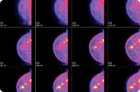
Breast scintigraphy is a nuclear imaging technique that uses intravenous injection of a radiopharmaceutical to evaluate the tissue's functional activity rather than its anatomy.
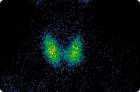
Parathyroid scintigraphy is a nuclear medicine test that helps find an overactive parathyroid gland, which is a gland that produces too much parathyroid hormone.
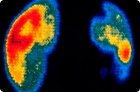
Renal scintigraphy is a nuclear medicine procedure in which a radiopharmaceutical is injected into a vein, and a gamma camera then records how the drug passes through the kidneys, accumulates in tissue, and is excreted in the urine.
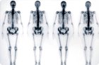
Bone scintigraphy, or osteoscintigraphy, is a nuclear medicine technique in which a small amount of a radiopharmaceutical is injected into a vein, and then a special gamma camera records its distribution in bone tissue.
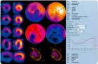
Myocardial scintigraphy is a nuclear cardiology technique that uses a small amount of radiopharmaceutical to evaluate how well blood flows to the heart muscle at rest and under stress.
A radionuclide is an unstable isotope that becomes more stable when it releases energy as radiation (nuclear decay). This radiation may include the emission of macroparticles or gamma-ray photons.
Imaging, or radiological, methods play a key role in the diagnosis and differential diagnosis of kidney diseases. Their role has increased significantly in recent years due to technical advances, which have significantly increased their resolution and safety.