^

Health

MRI diagnostics

MRI of the temporomandibular joint

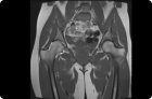
MRI of the temporomandibular joint is a promising method for diagnosing disorders of the motor function of the cranial bones. It allows you to quickly, without violating the integrity of soft tissues, assess the anatomical features and possible damage to the bones of the joint, its innervation, the state of the facial muscles, providing the doctor with important information for making an accurate diagnosis.

MRI of the brain with contrast

Magnetic resonance imaging of the brain using a contrast agent has a wide range of applications. But many patients are wary of intravenous / oral contrast because of the risk of complications.

MRI sacrum and coccyx: how is the procedure?

The MRI does not use light rays, it is based on the properties of hydrogen atoms to react to a magnetic field, and after the effect of the field ceases, everything returns to normal.

MRI of brain, head and neck vessels with and without contrast

Magnetic resonance imaging of cerebral vessels is the study of vascular structures using nuclear magnetic resonance. Allows you to assess the bloodstream of the brain and identify its pathology, damage.

MRI of the cervical spine with or without contrast: indications, technique

Modern diagnostic studies based on recording the electromagnetic response of atomic nuclei, which consist of the structures of the internal organs of the human body (magnetic resonance imaging), are in many cases the most informative noninvasive variants of visualization of pathologies invisible at external examination

MRI of the thoracic spine

From the standpoint of evidence-based medicine, the findings of this high-tech clinical study have one of the highest levels of reliability of results.

MRI of the small pelvis with and without contrast: preparation, what shows

One of the informative methods that will always help to establish an accurate diagnosis is the pelvic floor, widely used for diagnostic purposes in a variety of diseases.

MRI of the foot

The use of foot in the instrumental diagnosis of MRI allows orthopedists and traumatologists to accurately detect any diseases and detect traumatic injuries, degenerative changes or congenital anomalies of bones, joints and soft tissues of all parts of the foot.

MRI of the wrist joint

The wrist joint has many features: despite its small size, this articulation is subjected to constant and significant loads.

MRI of the joints of the hands

Magnetic resonance imaging of extremities allows you to carefully diagnose the condition of bone and soft tissues, joints. MRI provides a three-dimensional image.

Pages

You are reporting a typo in the following text:
Simply click the "Send typo report" button to complete the report. You can also include a comment.