Medical expert of the article

New publications
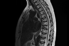
MRI of the thoracic spine
Last reviewed: 04.07.2025

All iLive content is medically reviewed or fact checked to ensure as much factual accuracy as possible.
We have strict sourcing guidelines and only link to reputable media sites, academic research institutions and, whenever possible, medically peer reviewed studies. Note that the numbers in parentheses ([1], [2], etc.) are clickable links to these studies.
If you feel that any of our content is inaccurate, out-of-date, or otherwise questionable, please select it and press Ctrl + Enter.
Today it is difficult to imagine traumatology, vertebrology, surgery, neurology or oncology without such a hardware (instrumental) diagnostic method as MRI of the thoracic spine.
From the standpoint of evidence-based medicine, the data from this high-tech clinical study have one of the highest levels of reliability of results.
 [
[ Indications for the procedure
First of all, conducting an MRI of the thoracic spine, as well as a simultaneous MRI of the cervicothoracic spine - when patients complain of pain in the chest and the corresponding part of the spinal column - is indicated to determine their causes.
Among the most probable diagnoses established after this examination of the bone structures of the spinal column and adjacent soft tissues are:
- violation of the integrity or displacement of vertebrae due to injuries to the thoracic (cervicothoracic) spine;
- protrusions, hernias or ruptures of intervertebral discs;
- damage to the ligamentous apparatus (interspinous and supraspinous ligaments);
- scoliosis and congenital spinal deformities (kyphosis, including juvenile, spondylolysis, etc.);
- degenerative-dystrophic pathologies (osteochondrosis);
- inflammation associated with osteomyelitis or spondylitis (including tuberculosis);
- radicular (or neurovascular) compression syndromes;
- intercostal neuralgia;
- cystic and tumor formations, spinal cord cancer.
What does an MRI of the thoracic spine show?
Three-dimensional images (layer-by-layer, in any planes) of all 12 vertebrae of this section (vertebrae thoracales T1-T12) - with intervertebral discs, facet joints, ligaments and tendons, foraminal openings (with blood vessels and nerve roots that come out of them), as well as the vertebral (vertebral or spinal) canal - with the arches and bony processes of the vertebrae that form it and the spinal cord located in it.
How long does an MRI of the thoracic spine take? The time required to conduct this examination does not exceed 25-30 minutes.
What does an MRI of the chest show? This examination visualizes all organs and anatomical structures of the thoracic cavity: trachea and esophagus; lungs, bronchi and pleural cavity; all sections of the mediastinum; heart (with its chambers, valves and vessels); sternum, ribs and intercostal muscles; thyroid and thymus glands, as well as the network of blood vessels, lymphatic vessels and nodes.
Answers to all questions of interest to patients: what is the preparation for MRI of the thoracic spine, the technique of conducting an examination on tunnel-type tomographs, contraindications for the procedure, possible consequences and complications after the procedure, as well as care after the procedure, are described in detail in the publication - MRI (magnetic resonance imaging)
And about how the MRI conclusion of the thoracic spine is deciphered (based on the obtained tomograms), you can find out in the material - MRI of the spine