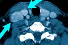
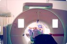
A computed tomography (CT) scan of the lungs is a cross-sectional imaging technique that uses an X-ray tube and detectors to rotate around the body, and a computer to assemble many thin slices into a detailed image of the lungs, pleura, mediastinum, great vessels, chest bones, and parts of the upper abdomen.