^

Health

CT scan

Magnetic resonance angiography

Magnetic resonance angiography (MP-angiography), in contrast to spiral CT, conventional and digital subtraction angiography, allows visualization of blood vessels even without the use of contrast media. The study can be carried out in 2D or 3D modes.

Magnetic resonance imaging (MRI) of the brain

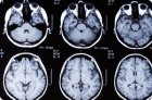
MRI examines the state of brain structures according to their shape, size and tissue density. It should be taken into account that MRI reflects the density of tissues depending on the water content in them, and therefore, first of all, such lesions as swelling-swelling of the brain (ONGM), demyelinating diseases, tumors are revealed.

CT-myelosysternoraphy

CT-myelosysternoraphy is a method that combines the possibilities of CT and myelography. Refers to invasive methods of imaging, because it requires the introduction of a sub-abdominal space contrast medium.

Tomography of the maxillofacial region

Tomography of the maxillofacial region is used in case of difficulties in evaluating the summation image in conventional pictures. These difficulties can be caused, in particular, by the complex anatomical structure of the maxillofacial region.

CT angiography

CT angiographic images need to be analyzed in different MIP projections (maximum intensity projections), MPR (multiplanar reconstruction), or 3D VRT reconstruction (volumetric imaging method).

Computed tomography of the abdominal cavity

All parenchymal organs should be seen equally well. An exception can be only the manifestation of the effect of a particular volume and the early arterial phase of contrast enhancement in spiral scanning. Such structures as blood vessels and intestinal loops should also be clearly visualized against the background of fatty tissue. The same applies to muscles.

Computed tomography of the chest

As a rule, the thoracic cavity is examined in the transverse direction (axial sections) with a cut thickness and a scanning step of 8-10 mm. For example, when making cuts with a thickness of 10 mm, with an overlap of 1 mm, table advance is carried out with a pitch of 8 mm.

Computer tomography of the neck

If there are no contraindications, computed tomography of the neck is performed after intravenous administration of contrast medium. The use of contrast preparations allows to determine more reliably the presence of malignant neoplasm and inflammatory process. To adequately enhance the vessels of the neck, more contrast material is required than, for example, for computed tomography of the head.

Head pathology on computed tomography

A direct consequence of a trauma to the skull is a brain contusion accompanied by a hemorrhage. Acute hemorrhage looks like a zone of increased density with edema of surrounding tissues and displacement of nearby brain structures.

Computer tomography of the head is normal

Scanning usually starts from the base of the skull and continues upwards. The resulting images on the film are oriented so that the slices are visible from the caudal side (bottom). Therefore, all the anatomical structures turn upside down from left to right. The topogram shows the location of each section.

Pages

You are reporting a typo in the following text:
Simply click the "Send typo report" button to complete the report. You can also include a comment.