Medical expert of the article

New publications
MRI of the temporomandibular joint
Last reviewed: 04.07.2025

All iLive content is medically reviewed or fact checked to ensure as much factual accuracy as possible.
We have strict sourcing guidelines and only link to reputable media sites, academic research institutions and, whenever possible, medically peer reviewed studies. Note that the numbers in parentheses ([1], [2], etc.) are clickable links to these studies.
If you feel that any of our content is inaccurate, out-of-date, or otherwise questionable, please select it and press Ctrl + Enter.
MRI of the temporomandibular joint is a promising method for diagnosing disorders of the motor function of the cranial bones. It allows one to quickly, without violating the integrity of soft tissues, assess the anatomical features and possible damage to the bones of the joint, its innervation, the condition of the facial muscles, providing the doctor with important information for making an accurate diagnosis.
Indications for the procedure
As we know from human anatomy, the lower jaw is the only movable bone of the facial skeleton, thanks to which we can take in and grind food, providing the body with the needs for nutrients. The mobility of the lower jaw is due to the temporomandibular joint, which is considered one of the most complex in the entire skeleton.
This joint is paired, and the movement of both its articulations must be synchronous and coordinated. But this movement is not simple, but combined, combining the sliding of the joint surfaces and their rotation around the floating center.
Sometimes, for various reasons, the coordinated work of the joint is disrupted, and in order to identify the cause of the disorders, doctors prescribe MRI of the temporomandibular joint, as one of the most informative methods.
Such examination is prescribed during a doctor's appointment, where the patient may come with various complaints. The following will be indications for MRI:
- unpleasant and painful sensations in the temples, cheek area, severe headaches,
- muscle tension in the lower jaw and cheekbones, neck, head,
- impaired sensitivity of the skin in the joint area,
- the appearance of a clicking sound (crunching) when moving the jaw,
- limited movement of the lower jaw, inability to open the mouth normally, speech and bite disorders,
- involuntary fixation of the jaw in one position, the inability to move it, open or close the mouth,
- difficulty chewing, discomfort while eating,
- rapid fatigue of the facial muscles,
- facial asymmetry,
- toothache after sleep,
- pain that pierces the jaw, neck and radiates to the shoulder,
- facial swelling not associated with lack of sleep, heart or kidney disease, unilateral facial swelling.
It is clear that MRI may be prescribed if there is a suspicion of dislocation or fracture of the jaw near the joint. The study is necessary both for diagnosis and for developing a treatment plan.
MRI diagnostics are prescribed if there is a suspicion of inflammatory processes in the joint (arthritis), rupture or perforation of the articular disc, osteoarthritis, osteomyelitis, synovitis, tumor processes in hard and soft tissues near the joint.
MRI of the temporomandibular joint is a preliminary (diagnostic) stage of orthodontic treatment and dental prosthetics.
Since MRI allows one to see details that are not accessible to other diagnostic methods (X-ray, orthopantogram, computer scanning), it is prescribed when the conducted study cannot detect the cause of the clinical symptoms. [ 1 ]
Preparation
Magnetic resonance imaging is a generally safe diagnostic method that allows you to obtain a lot of information about the internal structures of the skull skeleton without harming the patient's health. This examination does not require special preparation and can be carried out immediately upon arrival at the medical center.
Since MRI of the temporomandibular joint is prescribed by a doctor, the consultation with a specialist can be considered as preparation for the procedure. The doctor explains to the patient how safe the examination is, how it is carried out, what should not be taken into the machine, how to behave while the equipment is working so that the images are reliable and of high quality, what contraindications there are to MRI.
If the hospital doctor does not provide this information, the patient will learn all the information from the center staff who will perform the procedure.
If a person has a fear of enclosed spaces, they will be recommended options that will help them calm down and endure the procedure to the end. This may include taking sedatives or listening to music, for which special equipment is provided. In case of severe pain, painkillers are administered.
There are no restrictions on food, water consumption or medications. If the examination is planned to be carried out with contrast, which is rare, the patient will be told in advance how to prepare for it.
Technique MRI of the temporomandibular joint
The procedure for performing an MRI of the temporomandibular joint is extremely simple. The patient is asked to remove their outer clothing. You can take care of comfortable clothing in advance or use the one offered at the center. If you remain in your clothes, you will have to remove everything that contains metal parts (belts, buttons, snaps, cufflinks, dentures, etc.). Along with your outer clothing, you will have to leave behind all items that can interact with the magnetic field: watches, mobile phones, keys, payment cards, metal jewelry, etc.
In the room where the MRI equipment is located, the patient lies down on his back on a movable table, which will subsequently move in a magnetic circuit. At this time, the device takes several series of pictures. The patient's head is fixed with rollers, because immobility is the main condition for obtaining clear pictures, and not everyone can lie still for 5-15 minutes.
During the procedure, the patient has the opportunity to contact the doctor, who is in the next room. Two-way communication allows you to report discomfort and hear the doctor's instructions (for example, if necessary, hold your breath) in conditions of being in different rooms.
If the patient complains of limited mobility of the lower jaw, up to its jamming, an MRI of the left and right temporomandibular joint is performed, for which separate round radiofrequency coils are applied to them. Since the joint is paired, it is necessary to examine both of its sections, otherwise it is difficult to determine on which side the problem lies if the patient himself cannot indicate the localization of unpleasant sensations.
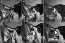
When clinical symptoms associated with jaw movement (during chewing, talking, opening the mouth, etc.) appear, an MRI of the temporomandibular joint with functional tests is prescribed. The two-phase study includes scanning with the mouth open and closed. Scanning with the mouth open is carried out for 5 minutes; to facilitate the fixation of the jaw, the patient is asked to clamp an object (usually a tube of toothpaste) between the teeth.
What does MRI of the temporomandibular joint show? On the images obtained using magnetic resonance imaging, it is possible to see microscopic structures with the possibility of examining them in different projections, to assess the condition of the mobile bone, articular disc, facial muscles and ligaments.
A tomogram makes it possible to examine various anatomical structures and changes in them, to identify foreign inclusions, tumor processes, congenital defects of the jaw and joint, and traumatic disorders. The doctor has the opportunity to assess the condition of the vessels in the area being examined, differentiate functional disorders and degenerative processes, and identify malocclusion and the causes that cause it. [ 2 ]
Contraindications to the procedure
MRI is considered one of the safest diagnostic methods, so it does not have a large list of contraindications that many other methods of examining the body have. Even existing contraindications are associated only with the possibility of interaction between the magnetic field and ferromagnets used in the manufacture of various devices. This interaction can damage the device and distort the images obtained by the tonometer.
There are very few absolute contraindications to MRI of the temporomandibular joint. This includes situations when a person, for medical reasons or for other reasons, cannot part with the device or metal objects in his body, i.e. the presence of
- pacemakers and defibrillators,
- inner ear implants (if they contain ferromagnets or the material of the device is unknown),
- vascular steel clips on aneurysm,
- any metal fragments, bullets in the human body, including small metal shavings inside the eye socket (professional reason requiring a preliminary X-ray of the employee's skull).
Relative contraindications are those related to the patient's condition and the presence of devices and appliances that should not change their properties under the influence of a magnetic field or somehow affect it. These include artificial joints, neurostimulators, insulin pumps, cardiac pacemakers, hemostatic clips and vascular stents, non-ferromagnetic implants. Questions may also arise regarding dental prostheses, steel plates and spokes used in orthopedic treatment, permanent makeup, tattoos, where ferromagnetic materials may be used.
In all of the above cases, the doctor must be informed in advance, if possible indicating what metal the device is made of, what makeup and tattoo paints were used, i.e. any information that will help to make the procedure safe and its results as reliable as possible.
MRI of the temporomandibular joint is not performed in cases of severe claustrophobia and serious condition of the patient, when life support procedures are required. During pregnancy, possible risks should be discussed with the doctor, although in general they are small, given the safety of a magnetic field of such strength for a person and the distance of the fetus from the examined area of the body (head). [ 3 ]
Care after the procedure
Considering the safety of the magnetic field and the tomograph itself, no consequences should be expected after the procedure. MRI diagnostics have become so popular because, in the absence of side effects during and after the procedure, it allows identifying pathologies that are inaccessible to other diagnostic methods. The only unpleasant consequence may be a series of poor-quality images if the patient did not remain still or hid the presence of metal objects in the body.
Complications after the procedure are also possible only if the patient does not listen to the doctor's warnings. There are few contraindications to the examination, but they must be taken into account. The magnetic field can disable the device, which may support the functionality of individual organs. For example, if the pacemaker malfunctions, the heart function is disrupted, which can lead to a serious condition and even death of the patient.
On the other hand, any piece of ferromagnetic material can affect the magnetic field, distorting the results of the studies. If the doctor relies on them, there is a risk of making an incorrect diagnosis and treatment that does not correspond to the situation.
No specific care is required after the MRI procedure of the temporomandibular joint. The main thing is that the results of the study help to identify the problem, and subsequently return the person's health and joy of existence.