Medical expert of the article

New publications
Cardiovascular MRI
Last reviewed: 29.06.2025

All iLive content is medically reviewed or fact checked to ensure as much factual accuracy as possible.
We have strict sourcing guidelines and only link to reputable media sites, academic research institutions and, whenever possible, medically peer reviewed studies. Note that the numbers in parentheses ([1], [2], etc.) are clickable links to these studies.
If you feel that any of our content is inaccurate, out-of-date, or otherwise questionable, please select it and press Ctrl + Enter.
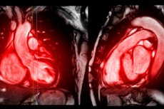
MRI (magnetic resonance imaging) of the heart and blood vessels is a highly accurate, non-invasive diagnostic method that provides detailed images of the heart and blood vessels without the use of ionizing radiation, unlike traditional X-rays and CT scans.
Using a powerful magnetic field and radio waves, cardiac and vascular MRI creates detailed images that can show the structure and function of the cardiovascular system, including the anatomy of the heart, the thickness and movement of its walls, the condition of valves and large vessels such as the aorta, venous and arterial vessels. This method can detect abnormalities such as heart defects, aneurysms, vascular blockages, cardiomyopathies, thrombosis and other heart and vascular diseases.
Benefits of cardiac and vascular MRI:
- High accuracy and contrast of images: MRI provides high tissue detail, which allows for accurate diagnosis of various pathologies.
- No ionizing radiation: Unlike CT scans and X-rays, MRI does not use ionizing radiation, making the procedure safer, especially for repeat examinations.
- Ability to obtain images in different planes: MRI allows visualization of heart and vascular structures in three dimensions, which simplifies the diagnosis of complex anomalies.
- Functional diagnosis: MRI can assess not only anatomy but also heart and vascular function, such as blood flow through different parts of the heart and vessels, and valve function.
Cardiac and vascular MRI may require the use of a contrast agent, usually gadolinium-based, to enhance visualization of certain structures or pathologies.
This method is widely used in cardiology and angiology for comprehensive cardiovascular evaluation and treatment planning.
Indications for the procedure
Cardiac MRI is used to diagnose various diseases and conditions of the cardiovascular system. Here are the main indications for cardiac MRI:
- Assessment of cardiac anatomy and function: to examine in detail the structure of the heart chambers, myocardial wall thickness and motion, and to evaluate heart valve function.
- Congenital (congenital) heart defects: Cardiac MRI can be used to evaluate anatomic features and plan surgical intervention in patients with congenital heart defects.
- Cardiomyopathies: for the diagnosis of various forms of cardiomyopathies, including hypertrophic, dilated and restrictive cardiomyopathies, and for assessing the extent of myocardial damage.
- Heart valve disease: to evaluate the structure and function of heart valves, including valve stenosis and insufficiency.
- Ischemic heart disease and assessment of myocardial viability: Cardiac MRI can be used to identify areas of myocardium with compromised blood supply and to assess the potential for cardiac tissue recovery after treatment.
- Cardiac and pericardial tumors: to detect and evaluate the nature of cardiac and pericardial tumors, including primary and metastatic tumors.
- Pericardial diseases: including pericarditis, pericardial effusion and constrictive pericarditis.
- AorticAneurysms and Dissection: for the diagnosis and evaluation of aortic aneurysms and dissections as well as other large vessels.
- Thrombosis and venous abnormalities: including thrombosis in the cavities of the heart, and venous outflow abnormalities.
- Monitoring and evaluation of treatment effectiveness: Cardiac MRI can be used to monitor changes in cardiac structure and function in response to drug or surgical treatment.
This list of indications is not exhaustive, and the possibility of cardiac MRI should be discussed with the attending physician based on the individual characteristics of the patient's condition.
Technique MRI of the heart and blood vessels
Cardiac MRI is an advanced and highly accurate imaging technique that assesses the anatomy, function and structure of the heart with a high degree of accuracy. This technique is particularly important for the diagnosis and treatment of heart disease, including congenital heart disease, ischemic heart disease, cardiomyopathies and pericardial disease. Here are some key aspects of cardiac MRI techniques:
- Cardiac MRI Technique: Cardiac MRI provides 3D analysis of global and regional cardiac function with high accuracy and reproducibility. There are various approaches to reconstruct cardiac motion and analyze cardiac deformation from MR image sequences, including cinematic MRI, tagged MRI, phase-contrast MRI, DENSE and SENC (Wang & Amini, 2012).
- Cardiac dynamics: MRI allows non-invasive assessment of cardiac biomechanical dynamics by providing tomographic images of the heart during different phases of the cardiac cycle, which is used to assess global cardiac function and regional endocardial motion. In addition, MRI can provide detailed information on motion patterns within the heart wall (Axel, 2002).
- Quantification: Cardiac MRI offers several capture techniques for accurate and highly reproducible assessment of global and regional ventricular function, flow, and perfusion at rest and during pharmacologic or exercise stress. Despite advances in hardware and software, quantitative image analysis often still requires manual contouring, which limits the clinical application of cardiac MRI (van der Geest & Reiber, 1999).
These basic aspects of cardiac MRI technique emphasize its importance and complexity as a tool for diagnosing and monitoring cardiac disease.
Contraindications to the procedure
The study of contraindications to cardiac MRI allows to identify a number of conditions under which this procedure may be dangerous or inapplicable for the patient. It is important to keep in mind that cardiac MRI is a highly accurate diagnostic method that requires a specialized approach depending on the patient's condition and the presence of certain risk factors. Based on general medical imaging knowledge and recommendations, there are several key contraindications to cardiac MRI:
- Thepresence of metal implants or fragments in the body, such as pacemakers, defibrillators, certain types of artificial heart valves, metal brackets or fasteners. The magnetic field of MRI can affect these devices, causing them to shift or malfunction.
- Claustrophobia or inability to remain still for long periods of time. To obtain good quality images, the patient must lie still for the entire examination, which can be difficult in claustrophobic patients without the use of sedation.
- Severe condition of the patient when transportation to the MRI and being in the machine is life-threatening.
- Presence of electronic hearing implants. MRI may damage the functioning of these devices.
- Pregnancy, especially in the early stages. Although MRI is considered a safe procedure, in early pregnancy it is preferable to avoid any exposure unless absolutely necessary.
These contraindications may vary depending on the type of MRI machine, its power and the specifics of the study. It is always important to consult with your physician and MRI specialists beforehand to assess all potential risks and contraindications in an individual case.
Normal performance
Normal cardiac MRI values include a number of parameters that reflect the structure and function of the heart and blood vessels. It is important to realize that the exact normal values may vary depending on the age, gender and individual characteristics of the patient, as well as the techniques and equipment used in a particular laboratory. The following are general parameters that are commonly evaluated as part of a cardiac MRI:
- Dimensions of the cardiac chambers:
- Left ventricle: normal size in diastole and systole.
- Right ventricle: normal size in diastole.
- Atria: absence of dilatation.
- Cardiac wall thickness:
- Left ventricle: normal myocardial thickness in diastole is usually 6-11 mm.
- Right ventricle: wall thickness is usually less than that of the left ventricle.
- Ventricular function:
- Left ventricular (LV) ejection fraction: normal values are 55-70%.
- Right ventricular (RV) ejection fraction: normal values are similar to LV.
- Left ventricular myocardial mass: within normal limits for the patient's age and sex.
- Myocardial status: no evidence of fibrosis or sclerosis that can be detected by assessment with delayed contrast sequences.
- Heart valve status: no significant regurgitation (backflow) or stenosis (narrowing).
- Anatomy and condition of the aorta and other large vessels: absence of aneurysms, dissections and narrowings.
- Blood flow through the vessels and valves of the heart: normalblood flow, with no evidence of obstruction or pathologic shunting.
- Pericardium: absence of thickening and effusion.
These parameters can be used by physicians to evaluate the patient's cardiovascular system and to detect various pathologies. It is important that the interpretation of cardiac MRI results be performed by a qualified specialist, as some measurements may require comparison with normative data specific to a particular laboratory or center.
Complications after the procedure
Magnetic resonance imaging (MRI) of the heart is a safe and non-invasive diagnostic technique used to evaluate the structure and function of the heart. Complications following cardiac MRI are extremely rare, especially when compared to other medical procedures involving the administration of contrast agents or the use of ionizing radiation. However, some risks and complications can occur:
- Allergic reaction to contrast agent: Although the gadolinium-based contrast agents used in MRI are considered safe, in rare cases they may cause an allergic reaction. Symptoms may include skin rash, itching, shortness of breath, or swelling of the face and throat. It is important to tell medical staff about any known allergies in advance.
- Nephrogenic systemic fibrosis (NSF): This is a serious complication associated with the use of gadolinium contrast agents in patients with severe renal dysfunction. NSF can lead to skin thickening, restricted movement, and even kidney failure. The risk of NSF is reduced by careful patient selection and avoiding the use of gadolinium contrast agents in individuals with severe renal impairment.
- Claustrophobia and discomfort: Some people may experience discomfort or claustrophobia due to the need to lie in the cramped space of the MRI machine for long periods of time. In such cases, the use of sedation may be necessary.
- Sedationside effects: If sedation has been used to reduce anxiety or claustrophobia, side effects such as dizziness, nausea, or drowsiness may occur.
In general, cardiac MRI is considered a safe procedure, and complications from the procedure are extremely rare. To minimize risks, it is important to carefully follow the instructions of medical personnel and provide complete medical information before the procedure. If you experience any symptoms or complications after a cardiac MRI, you should seek medical attention immediately.
Care after the procedure
Care after a cardiac magnetic resonance imaging (MRI) procedure does not require special measures in most cases, as MRI is a painless and safe procedure that does not involve exposure to ionizing radiation. However, there are general recommendations and precautions to consider:
- Return to normal activities: After cardiac MRI, patients can usually return to their normal activities immediately, unless otherwise advised by their physician. The procedure does not require a recovery period.
- Following the doctor's instructions: If contrast solution has been used for an MRI, it is important to monitor your body's reaction and seek immediate medical attention if you experience unusual symptoms (such as allergic reactions).
- Fluid Intake: If contrast is used, it may be recommended to increase fluid intake during the day after the procedure to allow for more efficient elimination of the contrast agent from the body.
- Health monitoring: If any unexpected symptoms or side effects occur after an MRI, you should contact your doctor immediately.
- Obtaining and discussing the results: The results of an MRI scan of the heart are usually available within a few days of the test. It is important to discuss them with your doctor to understand your medical condition and the need for further treatment or monitoring.
- Adherence to treatment recommendations: If a cardiac MRI has been performed as part of the diagnosis of a specific disease, the doctor's prescriptions and recommendations for treatment and follow-up care should be strictly followed.
Cardiac MRI does not affect the patient's physical condition and does not require special recovery, but it is important to pay close attention to any recommendations from a medical professional and report any changes in health.