New publications
Artificial intelligence tool reveals sex differences in brain structure
Last reviewed: 02.07.2025

All iLive content is medically reviewed or fact checked to ensure as much factual accuracy as possible.
We have strict sourcing guidelines and only link to reputable media sites, academic research institutions and, whenever possible, medically peer reviewed studies. Note that the numbers in parentheses ([1], [2], etc.) are clickable links to these studies.
If you feel that any of our content is inaccurate, out-of-date, or otherwise questionable, please select it and press Ctrl + Enter.
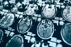
Artificial intelligence (AI) computer programs that process MRI scans reveal differences in the organization of the brains of men and women at the cellular level, a new study shows. These differences were found in white matter, the tissue found mostly in the inner layer of the human brain that facilitates communication between regions.
Men and women are known to suffer differently from multiple sclerosis, autism spectrum disorder, migraines and other brain problems, and to exhibit different symptoms. A detailed understanding of how biological sex affects the brain is seen as a way to improve diagnostic tools and treatments. However, while the size, shape and weight of the brain have been studied, researchers have only a partial understanding of its structure at the cellular level.
A new study led by researchers at NYU Langone Health used an AI technique called machine learning to analyze thousands of MRI brain scans from 471 men and 560 women. The results showed that the computer programs could accurately differentiate between male and female brains, identifying structural and complex patterns that were invisible to the human eye.
The results were confirmed by three different AI models designed to determine biological sex, using their relative strengths in either focusing on small patches of white matter or analyzing connections across large areas of the brain.
"Our findings provide a clearer picture of the structure of the living human brain, which may offer new insights into how many psychiatric and neurological disorders develop and why they may manifest differently in men and women," said lead study author and neuroradiologist Yvonne Lui, MD.
Lui, a professor and vice chair for research in the department of radiology at NYU Grossman School of Medicine, notes that previous studies of brain microstructure have relied heavily on animal models and human tissue samples. In addition, the validity of some of these past findings has been questioned by the use of statistical analyses of “hand-drawn” regions of interest, which required researchers to make many subjective decisions about the shape, size, and location of the regions they selected. Such choices could potentially skew the results, Lui says.
The new study's findings avoided this problem by using machine learning to analyze entire groups of images without telling the computer to look at a specific location, which helped eliminate human biases, the authors note.
For the study, the team started by feeding the AI programs existing data of example MRI brain scans of healthy men and women, along with the biological sex of each scan. Because these models were designed to use sophisticated statistical and mathematical methods to get “smarter” over time as they accumulated data, they eventually “learned” to discern biological sex on their own. Importantly, the programs were restricted from using overall brain size and shape for their determinations, Lui says.
According to the results, all the models correctly identified the gender of the scans 92% to 98% of the time. Several features in particular helped the machines make their conclusions, including how easily and in what direction water was able to move through the brain tissue.
"These findings highlight the importance of diversity when studying diseases that originate in the human brain," said study co-author Junbo Chen, MS, a doctoral student at NYU Tandon School of Engineering.
"If, as has been the case historically, men are used as the standard model for various disorders, researchers may miss critical insights," added study co-author Vara Lakshmi Bayanagari, MS, a graduate research student at NYU Tandon School of Engineering.
Bayanagari cautions that while the AI tools could report differences in brain cell organization, they could not identify which gender was more prone to which traits. She adds that the study classified gender based on genetic information and included only MRI scans of cisgender men and women.
The team plans to further study the development of sex differences in brain structure over time to better understand the role of environmental, hormonal and social factors in these changes, the authors say.
The work was published in the journal Scientific Reports.