New publications
Serine against "diabetic" vessels in the retina: what the study showed
Last reviewed: 09.08.2025

All iLive content is medically reviewed or fact checked to ensure as much factual accuracy as possible.
We have strict sourcing guidelines and only link to reputable media sites, academic research institutions and, whenever possible, medically peer reviewed studies. Note that the numbers in parentheses ([1], [2], etc.) are clickable links to these studies.
If you feel that any of our content is inaccurate, out-of-date, or otherwise questionable, please select it and press Ctrl + Enter.
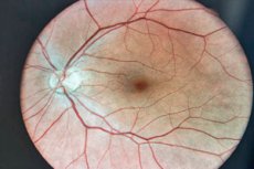
A paper published in the journal Theranostics by a group from Harvard/Children's Hospital Boston found that supplementation with the common amino acid serine significantly suppressed abnormal proliferation of blood vessels in the retina (neovascularization) in a classic mouse model of hypoxic retinopathy. This "abnormal" vascular formation underlies retinopathy of prematurity and proliferative diabetic retinopathy, two leading causes of vision loss.
The idea in a nutshell
During hypoxia, photoreceptors experience energy starvation and send a signal to "build more vessels" - this results in many fragile, leaky capillaries. The authors tested whether this pathological response would be weakened if the retina was fed with serine (a key amino acid in the metabolism of single-carbon groups and a precursor to a number of lipids). The answer is yes, and quite convincingly.
What exactly did they do?
- An oxygen-induced retinopathy (OIR) model was used: newborn mice were kept in 75% O₂ and then transferred to air - this causes a “wave-like” death and then retinal hypoxia with a peak of neovascularization on the 17th day of life.
- Serine was given systemically (intraperitoneally or orally) during a period of relative hypoxia. The mothers were separately placed on a diet low in serine/glycine to see the opposite effect.
- The groups were compared by the area of neovascularization and "bloodless" zones and a "multi-omic" analysis of the retina was performed: metabolomics, lipidomics, proteomics, scRNA-seq. Plus pharmacology: they blocked β-oxidation of fatty acids (ethomoxir/malonyl-CoA) and mitochondrial ATP synthase (oligomycin) to check what serine acts through.
Key Results
- Fewer pathological vessels. Serine significantly reduced the area of neovascularization; while serine/glycine deficiency in the mothers’ diet, on the contrary, increased it.
- Energy is the center of the story. The effect of serine disappeared when fat oxidation (FAO) or oxidative phosphorylation (OXPHOS) were inhibited. That is, the protection depends on mitochondria. In proteomics, there is an increase in OXPHOS proteins; in transcriptomics, there is an increase in “respiratory” genes and a decline in proangiogenic signals in the rod photoreceptor cluster.
- Lipid trace. Phosphatidylcholines, the most common class of membrane phospholipids, increased in the retina, which is logical for tissues with a colossal turnover of membranes (photoreceptors).
- Candidate mediator: HMGB1 has been identified as a possible nodal regulator through which serine dampens proangiogenic signals during hypoxia.
Why is this important?
Today’s “heavy” treatments — laser and anti-VEGF injections — save vision, but have limitations and potential risks, especially in infants. A simple nutritional strategy targeting retinal neuronal metabolism could be a gentle complement or “bridge” between treatments. Observational data in humans are indirectly consistent: low serine is associated with macular neovascularization, and serine/glycine pathway remodeling has been described in ROP and diabetic retinopathy. This work adds causality, albeit in a model.
Be careful: these are mice for now
- OIR is a model, not a complete copy of human diseases; direct “translation” of serine doses to humans cannot be done.
- Systemic amino acid supplementation is not a “harmless vitamin”: in some conditions, excess amino acids/metabolic shifts can have side effects.
- Clinical studies are needed: safety regimens, windows of efficacy (in preterm vs adults with diabetic retinopathy), combination with anti-VEGF and impact on baseline vascular remodeling.
What's next?
Logical next steps are small clinical pilots with mitochondrial function/retinal lipid profile biomarkers, testing serine in combination with existing therapies, and finding precise “molecular knobs” (same HMGB1) for targeted intervention without systemic amino acid loading.