Medical expert of the article

New publications
MRI of the foot
Last reviewed: 03.07.2025

All iLive content is medically reviewed or fact checked to ensure as much factual accuracy as possible.
We have strict sourcing guidelines and only link to reputable media sites, academic research institutions and, whenever possible, medically peer reviewed studies. Note that the numbers in parentheses ([1], [2], etc.) are clickable links to these studies.
If you feel that any of our content is inaccurate, out-of-date, or otherwise questionable, please select it and press Ctrl + Enter.
Indications for the procedure
Magnetic resonance imaging – MRI of the foot – is prescribed to patients with complaints of pain in the foot or ankle joint, joint stiffness and problems with walking. Visualization is performed to accurately determine pathological changes in anatomical structures, allowing to establish the true causes of pain syndrome, which can occur for a variety of reasons, in particular:
- in case of bone fractures;
- in complex cases of ligament sprains;
- due to damage (rupture) or enthesopathy of the Achilles tendon;
- if the joints of the interphalangeal joints of the fingers are inflamed (with arthritis and arthrosis) or their joint bags with the development of bursitis of the foot;
- due to deformation of joints and development of ankylosis;
- when the plantar fascia becomes inflamed, that is, with plantar fasciitis;
- if a periarticular tumor forms - hygroma of the foot;
- for inflammation of soft tissues (abscess, phlegmon, diabetic foot, gout ).
MRI of the heel of the foot is performed, first of all, in the case of the formation of a marginal osteophyte (heel spur), as well as in cases of inflammation of the heel bone (epiphysitis, osteonecrosis); damage or deformation of the heel (Achilles) tendon.
This diagnostic method is indispensable for choosing the optimal tactics for any surgical interventions, including those aimed at correcting congenital foot defects (peromelia, syndactyly, ectrodactyly, equine foot).
Preparation
Preparation for any MRI involves the patient removing all metal objects before the procedure begins.
 [ 6 ]
[ 6 ]
Technique MRI of the foot
When using a closed tomograph (tunnel type) or an open panoramic scanner, the patient assumes a horizontal position, the limbs are fixed, since complete immobility is important during scanning. There are tomograph models that allow the examination to be carried out when the patient is sitting.
The average duration of a foot MRI is half an hour. No post-procedure care is required.
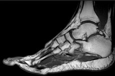
What does an MRI of the foot show?
Using three-dimensional images obtained using magnetic resonance imaging, doctors can clearly see existing changes and damage to bone structures.
MRI of the joints of the foot – subtalar, talocalcaneonavicular, calcaneocuboid, cuneonavicular, tarsometatarsal, intermetatarsal and interphalangeal joints – shows the condition of all joint structures. This concerns the joint capsule and its synovial membrane, the epiphyses of the joint-forming bones, the joint cavity and cartilage.
MRI of the soft tissues of the foot visualizes the fat pads of the sole, heel, toes and can show edema, foci of infiltration and inflammation. In addition, an image of all the muscles of the dorsum and plantar part of the foot, all tendons and tendon ligaments, blood vessels and nerves is obtained.
Although the images are saved on a computer and issued on film or electronic media, a radiologist (a specialist in tomographic diagnostics) makes a medical report, transcript or description of the MRI of the foot - indicating the existing pathological changes, their nature and exact localization.
Contraindications to the procedure
MRI scanning, including of the foot, is contraindicated in patients with: a pacemaker; a continuously delivering insulin device (insulin pump); cochlear implants; metal stents, surgical staples, pins, plates, screws, etc.
Contraindications also include the first trimester of pregnancy; inability to remain completely still for at least 30 minutes; the presence of such a psychopathic syndrome as claustrophobia.
Complications after the procedure
MRI uses radiofrequency pulses that pass through an electromagnetic field, so there are no consequences associated with “irradiation” of the body, that is, exposure to ionizing radiation.
However, some patients – exclusively in cases where the electromagnetic field voltage is exceeded – may experience minor complications after the procedure, such as mild dizziness, short-term fasciculations (spontaneous twitching of individual muscle fibers) and a metallic taste in the mouth.
However, the majority of patients’ reviews of their well-being after magnetic resonance imaging do not contain any complaints.
Which is better, CT or MRI of the foot?
Considering the importance of MRI results of the foot for correct diagnosis and selection of optimal treatment tactics, this method of instrumental diagnostics is preferable to CT, as it visualizes the structures of the foot in different planes and with higher contrast (this is especially true for dense connective tissue of cartilage and ligaments).
In addition, unlike CT (which uses ionizing radiation), MRI is not an X-ray method, and the frequency of its use is not limited.