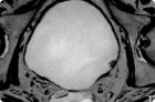
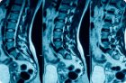
Magnetic resonance imaging of the bladder is a non-invasive imaging technique that allows detailed evaluation of the bladder wall, surrounding tissue, and adjacent organs without ionizing radiation.
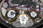
Retroperitoneal magnetic resonance imaging (MRI) is a layered imaging technique that uses a powerful magnetic field and radio waves to produce detailed images of organs and tissues without ionizing radiation.
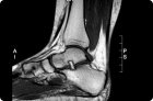
Ankle magnetic resonance imaging (MRI) is a noninvasive test that uses a strong magnetic field, radiofrequency pulses, and computer signal processing to produce layered images of the joint, ligaments, tendons, bones, cartilage, and bone marrow.
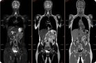
Whole-body MRI, or magnetic resonance imaging, is a non-invasive test that uses a strong magnetic field, radiofrequency pulses, and computer processing to produce images of multiple anatomical areas in a single visit.
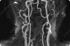
In clinical practice, the term "MRI of arteries" usually refers to magnetic resonance angiography, that is, the study of the arterial bed using a magnetic field, radiofrequency pulses, and computer signal processing.
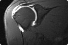
Arthrography is a method of visualizing a joint after the introduction of a contrast agent into the joint cavity or, less commonly, after the intravenous administration of contrast with its subsequent accumulation in the joint.
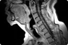
Magnetic resonance imaging (MRI) of the soft tissues of the neck is a layered imaging technique that uses a strong magnetic field, radio waves, and computer signal processing to produce detailed images of soft tissues.
Magnetic resonance imaging of bones is an imaging technique that uses a strong magnetic field, radiofrequency pulses, and computer processing to produce detailed images.
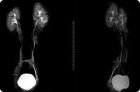
Magnetic resonance urography is a method of radiological diagnostics that allows for the evaluation of the kidneys, renal pelvis, ureters, and bladder in a single examination.