Medical expert of the article

New publications
Flat moles
Last reviewed: 04.07.2025

All iLive content is medically reviewed or fact checked to ensure as much factual accuracy as possible.
We have strict sourcing guidelines and only link to reputable media sites, academic research institutions and, whenever possible, medically peer reviewed studies. Note that the numbers in parentheses ([1], [2], etc.) are clickable links to these studies.
If you feel that any of our content is inaccurate, out-of-date, or otherwise questionable, please select it and press Ctrl + Enter.
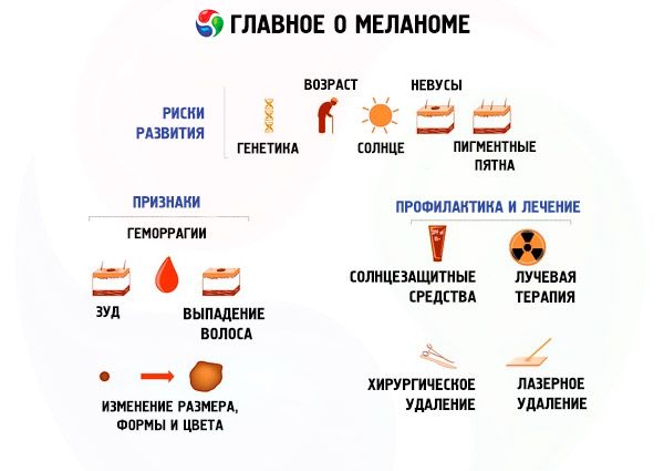
Everyone has moles, or nevi. They are benign growths of varying shades of brown. Their color comes from melanin, a pigment found in melanocytes (skin cells) that make up nevi.
They are considered small if they have a diameter of up to 15 mm; medium – up to 100 mm; large – more than 100 mm; giant – occupying significant areas of the skin of parts of the body.
 [ 1 ]
[ 1 ]
Causes flat moles
Lentigo are the most common flat moles formed by melanocytes. They are oval or round in shape, 3-5 mm in diameter, with smooth or raised edges. The number of moles is proportional to the time spent in the sun (solar lentigines).
Senile flat moles appear for the same reasons, with the onset of age-related skin changes. Usually, after 45 years, their number and size increase, the color becomes darker. Localization sites are the face, hands, shoulders and neck.
The cause of juvenile (simple) lentigo is not established, but it has nothing to do with sun exposure. Round or oval dark spots with smoothed or jagged edges appear in early childhood and in adolescents. They are found on the skin and mucous membranes, and can be single or scattered.
There are no such moles on the body of babies. They appear later. At first, a small flat spot forms on the skin, over time it can increase in size and rise above the skin layer. Whether the nevus will remain flat depends on the level of the pigment cells. If the melanocytes are located in the epidermis, the mole will be flat. If they are located deeper directly in the skin itself, it will be convex.
The formation of nevi and their number are influenced by heredity. Even the name "birthmark" speaks of this. They seem to be passed down by inheritance, sometimes even localized in the same places as in older relatives.
The formation of nevi can be facilitated by radiation and X-rays that affect the body from time to time. Viral and infectious diseases that leave marks on the skin, insect bites and other injuries can trigger the release of groups of melanocytes to the surface of the skin.
The intermediate lobe of the pituitary gland secretes a hormone that stimulates the production of melanocytes, so hormonal surges can cause nevi to appear. This may explain the appearance and disappearance of moles in teenagers and pregnant women.
Alternative medicine explains the formation of moles by the release of internal energy accumulating in the site of inflammation.
Symptoms flat moles
If a previously convex mole suddenly becomes flat, this may mean that the convex nevus was torn off and not noticed, and a flat spot of dark brown or black color appeared instead. This is an alarm bell, you should definitely contact an oncodermatologist and as soon as possible.
Clinically, the initial stage of melanoma resembles a flat dark nevus of an asymmetrical shape with jagged edges. These spots can have a diameter of 6 mm or more. A characteristic feature of melanoma is a shiny surface, brown or black.
It usually begins to grow noticeably, change color, can become bumpy and convex, crack, get wet, bleed. If such symptoms appear, you cannot delay.
Pathogenesis: Melanoma spreads over the surface of the skin, gradually rising, and inward, successively growing into the layers of the skin and underlying tissues.
This tumor is characterized by rapid metastasis, usually to nearby lymph nodes. It often metastasizes to the skin, and brown or black rashes appear in a scattering near the primary tumor.
Metastases reach various organs through the blood, but the most common is classical metastasis (to the lungs, liver, brain and adrenal glands). Metastases are often multiple and free melanin is present in blood and urine tests.
[ 2 ]
Flat mole on face
Such nevi are not very common on the face. These are clusters of pigment cells in the epidermis without a deep root of any shade of brown with a flat dry surface. Hairs do not grow on such moles. Their size does not exceed 1 cm. Their shape, size and number do not change over time.
Beige-brown spots are called lentigo. Their number increases significantly in some teenagers, sunbathers, and almost all elderly people. This is the most common type of mole, which does not pose any danger.
Flat birthmarks of large or medium size are often found on the face. They are considered melanoma-risk moles.
Nevus of Ota is a large, bluish-colored neoplasm.
Dubreuil's melanosis is a medium-sized, light-brown, irregularly shaped neoplasm that tends to darken and gradually increase in size over time.
Most giant moles that occupy large areas of the face are flat. Such nevi appear in childhood, their size grows with the growth of their "owner", their color is also varied.
There is no need to worry about every mole, you can't remove them all, and there is no point in doing so. Most of them do not pose any health risks. However, nevi on the face are constantly exposed to the sun, and are not always good from an aesthetic point of view.
Changes in a mole on the face are hard to miss. It is impossible to remove it at home, you can cause yourself irreparable harm. Even if you need to remove a seemingly safe mole for aesthetic reasons, you should definitely consult a dermatologist, since only a specialist can correctly determine the method of removing the neoplasm.
[ 3 ]
Flat moles have become raised
When a harmless, long-familiar, flat mole becomes convex, you need to immediately put everything aside and consult an oncodermatologist. You need to do the same if a flat mole grows, as if spreading in different directions, losing its symmetrical shape and becoming denser. Such activity may be a sign of melanoma.
In this case, every minute counts. The sooner you start treatment, the greater the chances of avoiding mortal danger.
Forms
Excess melanin in skin cells causes pigment nevi to appear, and its quantity determines their color. If a flat black mole has been on the skin for a long time, does not change, and there are other such moles, they are most likely safe.
A black nevus should raise concerns if it has recently appeared, as well as if an old neoplasm has turned black in whole or in part.
Flat brown moles are located anywhere on the skin or mucous membranes. Insolation does not affect these nevi, they do not change color, their number does not increase, there are usually not many of them on the body. Flat brown moles are usually slightly darker than freckles.
Pink melanocytic nevi are found in fair-haired and red-haired people with fair skin. These are common moles, but in people with this phenotype, the melanocytes produce a pink-red pigment rather than a brown one.
Lentigo are multiple flat brown nevi, their number can increase greatly in children and adolescents (juvenile lentiginosis), as well as under the influence of solar radiation at any age, increasing over the years.
In addition to lentigo, there are also Mongolian and coffee spots. Mongolian spot is a flat large single spot or a group of brownish-blue asymmetrical spots in the lumbar region. They disappear on their own at 12-15 years.
A coffee stain is a flat, light-coffee-colored spot. When there are 1-2 such spots on the body, it is not dangerous. If there are three or more, there is a possibility of neurofibromatosis. It makes sense to consult a neurologist.
A red flat mole is formed from damaged capillaries of the circulatory system. It looks like a flat or slightly convex neoplasm of red color with uneven edges.
There are two types of superficial capillary birthmarks: hemangioma and vascular malformation.
Hemangioma is a bright red neoplasm that appears on the skin in the first weeks of life and may rise slightly above the skin surface. It does not disappear over time. It is usually located in the face and neck area. It never becomes malignant.
Vascular malformation is a congenital pathology of blood vessels. Its varieties include bright port-wine stains, which remain for life, and a "stork bite", the result of pressure from the mother's pelvic bones on the baby's skull in the last months of gestation. This stain disappears on its own in infancy.
If a mole resembles a small vascular red dot, it is probably an senile angioma. They begin to appear after 30 years, and with age, their number increases. Senile angiomas consist of dilated vessels located close to the surface of the skin.
A flat mole with jagged edges that remains unchanged for a long time most likely does not pose a danger, especially if there are other similar formations on the body.
But if the borders of the mole were previously smooth and have changed recently, or if it is a newly appeared mole, then you should see an onco-dermatologist.
A flat, flesh-colored mole is most likely a normal, harmless mole of a non-saturated color.
A decrease in the pigmentation of a brown mole to a flesh-colored color is also a reason to bring it to the attention of a doctor.
Complications and consequences
Are flat moles dangerous? These growths are safe in themselves. But you shouldn't ignore them completely. Flat nevi should be small, uniform in color, and have a rounded, symmetrical shape.
It is advisable to regularly observe moles with a mirror in your hands, especially if there are many of them. Sometimes a mole degenerates into melanoma – a malignant neoplasm. The mechanisms that trigger this process are different. The main reason is sunburn. Even when using sun protection, it is better to avoid direct sunlight on the skin. Mole locations should be avoided during massage, do not direct a strong stream of shower at them, and avoid mechanical damage by clothing. Dermatologists believe that during puberty and pregnancy, the risk of nevi degenerating is significantly higher.
The appearance of multiple new birthmarks on the skin is also an alarming factor.
According to observations, in most cases (about 80%) malignant skin neoplasms are formed not from existing moles, but from normal skin cells. At the beginning of the process, a new, not quite ordinary mole can be noticed.
When an old mole degenerates, its appearance begins to change. And any change should cause concern. Darkening of the nevus, its itching, tingling, cracks, bleeding, asymmetry that has appeared or a dark rim around it - all these are reasons to contact an oncodermatologist.
What to do if you tear off a flat mole? Basically, a full or partial tearing off of a mole can end quite harmlessly. Instead of the old nevus, a new one will appear, also benign. The damaged one will close safely and will never bother you.
If a mole is injured, you need to stop the bleeding (using three percent hydrogen peroxide), disinfect the wound and apply a dry sterile bandage. It is advisable to place the completely torn off nevus in a vessel with saline solution to analyze the malignancy of the cells. The probability of a change in the cellular structure of a flat mole as a result of injury increases several times.
After treating the wound, you should immediately contact an oncodermatologist. After the examination, the doctor will give recommendations on how to treat the wound and whether to remove the remaining parts of the mole.
After removing a mole or its remnants, you need to follow the surgeon's recommendations without trying to speed up the healing process.
You cannot remove the remaining parts of the torn off mole yourself, cauterize them with celandine, do not cover the wound with adhesive tape or make compresses.
If any circumstances prevent you from visiting a doctor, observe the injured nevus or the site of injury. If pathological changes such as itching, rapid growth, cracking, bleeding, or color change occur, see a specialist immediately.
Diagnostics flat moles
It is impossible to independently determine what is hidden behind an ordinary-looking mole.
The dermatological ABCD rule helps the doctor determine the danger of neoplasms, where A is the asymmetry of the mole, B is the degree of unevenness of its border, C is the uniformity of color, and D is a diameter of at least 6 mm. However, this rule is not absolute; a subjective, incorrect assessment of the set of signs is possible.
If there is a suspicion of melanoma, tests are done: histopathological microscopy of a smear taken from the surface of the mole. Biopsy - the material for analysis is taken from an area of skin near the mole, but not from the mole itself, since it is dangerous to injure this neoplasm even for the sake of establishing a diagnosis.
Modern oncology uses instrumental diagnostics of melanoma and other malignant skin lesions. These include dermatoscopy, infrared spectroscopy, confocal laser microscopy, high-frequency ultrasound, optical coherence tomography, and fluorescent diagnostics.
The most promising of the non-traumatic diagnostics is considered to be epiluminescent dermatoscopy, which allows the doctor to examine the pigment spot in incident light with 10-fold magnification.
This diagnostic procedure can be performed using a computer to which a dermatoscope is attached. The resulting series of digital images of the nevus are saved and analyzed using diagnostic algorithms based on comparative characteristics of dermatoscopy of benign and malignant skin neoplasms. Such diagnostics do not injure the mole and allow avoiding unnecessary surgical interventions.
Based on many years of studying the morphology and dynamics of pigmented skin neoplasms, as well as establishing their dermatoscopic features, diagnostic algorithms were developed to compare melanocytic and non-melanocytic nevi. Thus, differential diagnostics was created for early detection of skin melanoma based on its inherent structural features, which are revealed by dermatoscopy.
Who to contact?
Treatment flat moles
Can flat moles be removed? This question worries many. Flat moles can be removed. But is it worth it? If a mole has started to change, is damaged, is regularly injured by clothing, then it is better to remove it. If it is benign and does not interfere, then there is no need to remove it.
You can get rid of a mole for aesthetic reasons, but only after a dermatologist's examination. The neoplasm must be examined, a diagnosis and method of removal established. All these procedures are preferably carried out in an oncology medical institution. A histopathological examination of the removed neoplasm must be carried out to ensure that there are no signs of degeneration.
Removing moles is contraindicated in acute infectious and inflammatory processes, exacerbation of chronic diseases, cardiovascular diseases, autoimmune, mental and oncological diseases, pregnancy.
Removal of flat moles
There are several methods for removing benign nevi.
Cryodestruction involves destruction by cold and subsequent death of neoplasm cells under the influence of low-temperature liquid nitrogen. This method is used to remove flat moles. Its disadvantage is the possibility of injury to healthy tissue and incomplete destruction of the mole (the procedure will have to be repeated). Cryodestruction may leave burns, scars and marks, a long healing period, sometimes about six months.
Unlike the previous method, laser removal of flat moles has serious advantages. During the procedure, there is no pain, no risk of infection, and healthy tissues around the mole are not injured, since the removal parameters are strictly observed. Laser removal methods do not leave scars, so they are recommended for removing nevi on the face and exposed parts of the body. With the help of a laser, you can remove birthmarks located in hard-to-reach areas of the body.
The laser evaporation procedure involves the action of a laser beam on the mole, evaporating the liquid component from the cells of its tissue layer by layer.
The disadvantage of this method is the impossibility of conducting a histopathological examination, since nothing remains of the removed neoplasm.
The method of cutting out flat moles with a laser knife leaves the possibility of analyzing the tissue of the removed mole, but there is a risk of thermal burns to the skin.
The laser knife cuts away the pigmented skin layer by layer down to the layer where there is not a single melanocyte.
After removal, the mole wound is treated with an antiseptic and covered with a sterile bandage. It is protected from sunlight and moisture. A few days later, a crust forms, which falls off after a couple of weeks.
Removal of flat moles with a Surgitron (radio wave knife), developed and tested in the USA, has the following advantages: fast and painless operation; removal of moles located in hard-to-reach areas of the body; rapid skin regeneration.
This method can be used to excise not only safe nevi, but also some malignant nevi.
This method involves contactless removal of neoplasm tissues with a surgitron, with simultaneous stopping of bleeding and disinfection, leaving no postoperative scars. Only nevus tissues are subject to the destructive action of the radio wave knife, and the healthy skin around it is not injured. The operation usually lasts up to 0.5 hours, its duration depends on the number and size of the neoplasms being removed.
In addition to the contraindications listed above, removal of nevi with a radio knife is not performed if the patient has a pacemaker.
Large melanoma-risk moles are removed surgically. The procedure is performed under local anesthesia, the nevus is removed with a scalpel along with a small area of adjacent skin. After the operation, a scar remains, which heals for several weeks and requires care.
After removing moles, it is recommended to: treat the wound with an antiseptic three times a day; refrain from prolonged exposure to the sun; do not visit solariums, swimming pools, baths, saunas; immediately consult a doctor if you injure the removal site or find a lump or change in skin color in the area of the removed nevus.
How to remove flat moles at home?
Removing moles at home is very dangerous. It is strictly forbidden to try to get rid of large or noticeably changing nevi. This can cause irreparable harm to health. Only a doctor can give recommendations for removing a mole after an examination.
Nowadays, there are many different natural remedies for removing skin defects – warts and moles. They are sold without a prescription, but as a result of their use, you can lose your health and even your life.
So, here are the medications that can remove skin defects.
Stefalin ointment for getting rid of moles and warts is being distributed on the Internet. It is positioned as a 100% natural product without chemical additives, consisting of "a special inflorescence of herbs and roots collected on high-mountain slopes in different places." According to its distributors, it removes any nevi, warts and papillomas, including those dangerous for melanoma.
It is not so much the effectiveness of this ointment that is questionable, but its safety. It is not sold in pharmacies because it is not licensed and therefore has not undergone the necessary control tests.
Pharmacies sell medications that popular wisdom attributes the ability to remove all sorts of minor skin defects, including moles. But these products are primarily intended for medical workers, not for the average citizen to self-medicate.
Solcoderm solution is used externally to eliminate benign superficial skin defects, such as warts, actinokeratosis, seborrheic keratosis, pointed condylomas and nevocellular nevus (after checking for benignity). The instructions for use of the drug note that this solution should be applied by a doctor or under his supervision. It is not recommended to use Solcoderm on your own.
Pharmacies sell various celandine-based preparations, for example, Mountain Celandine - an alcohol concentrate of celandine for lubricating warts and papillomas, with the addition of gentian, string, golden rhododendron and goose foot. The instructions say nothing about moles, but this usually does not stop fans of self-medication.
An even harsher drug, Superchistotel, has nothing to do with herbs. It is a mixture of alkalis. It is sold in a small bottle with an applicator for applying the drug to the skin. Its components cause the death of skin cells as a result of an alkali burn. Skin cells die from necrosis, the neoplasm darkens, dries up and falls off. A scar may remain, and in particular, the official instructions for the drug do not mention the removal of moles.
The above and similar remedies are unlikely to help get rid of moles, but they can cause significant harm to health. Compared to these drugs, folk remedies based on natural ingredients are more gentle. However, it should be taken into account that no folk remedy is recognized as effective by official medicine.
Folk remedies
Radically dealing with moles even with folk remedies is dangerous. The recipes below may be suitable for removing freckles or very light small flat moles. At home, for the sake of clean and white skin, herbal treatment was used, as well as other means that were used in the household.
The safest of them are:
- fresh celandine juice - apply it to the neoplasm three times a day (you can use Vaseline - it has an even more gentle effect and is easy to apply);
- apple cider vinegar - use one drop per day on the mole, the procedure lasts 5 days;
- lapis pencil - the active substance (silver nitrate) has a disinfecting, necrotic effect, smear the neoplasm once every 10 days for a month;
- garlic and lemon juice are dripped alternately onto the nevus for about half a minute every day for 2 weeks;
- you can wipe the mole with castor oil, dandelion juice or pineapple juice 2 times a day (whitening effect);
- mix honey and flaxseed oil and make a compress on the nevus for 2 or 3 minutes;
- garlic gruel is applied to the mole, sealed with a bandage for no more than 4 hours (the skin around the mole is lubricated with Vaseline), it can be repeated after 3 or 4 days;
- vinegar essence – drip 1 drop per day from a pipette onto the nevus for a month (lubricate the skin around the mole with petroleum jelly).
And the safest method is a conspiracy:
- on a grain ear - cut off a grain ear and touch the hated nevus with the cut, bury the ear in the ground with the cut side up, as soon as it rots - the mole will disappear;
- for apples - cut a ripe, beautiful apple in half and rub one half against the other over the nevus, fold the halves into a whole apple, tie it and bury it in the ground; when the fruit rots, the mole will disappear;
- on potatoes - cut the potato in half, immediately bury one half in the ground, rub the mole with the other half and bury it near the first, accordingly, the mole will disappear when the potato halves rot.
[ 4 ]
Consequences of mole removal
Removing a nevus at home can lead to the most negative consequences. If you assume that you can get rid of it safely, you can, after some time, face a deadly danger - melanoma. In the early stages, it is often confused with an ordinary flat mole. Therefore, before removing a nevus in any way, you must consult an oncodermatologist who will conduct a full diagnosis.
More information of the treatment
Prevention
Prevention of damage to moles is the absence of clothing and footwear that exert pressure or rubbing on the neoplasms. In order not to accidentally tear off a nevus, you need to cut and file your nails short, and not wear tight clothing and shoes.
When you do not want to deny yourself long, well-groomed nails, tight skirts and jeans, belts, stylish and elegant shoes, you need to worry about your safety and consult a specialist about removing nevi located in places of possible injury. In this way, you can prevent many significant troubles.
It is best to remove nevi for cosmetic purposes when the hormonal background is stable (in early childhood or adulthood). The autumn or winter season is more suitable for this procedure, when solar radiation is least intense.
Forecast
It is not difficult to accidentally injure a mole, with nails, tight clothing, a washcloth or a razor. Such an incident does not always lead to sad consequences, do not believe rumors and various scary stories. Injury to a nevus can simply lead to a banal infection.
However, a benign nevus can become malignant as a result of injury. If a mole is accidentally damaged, you should see a doctor immediately. Even if a malignant mole is damaged, timely treatment will have better results.
[ 7 ]