Medical expert of the article

New publications
Removal of moles by electrocoagulation
Last reviewed: 06.07.2025

All iLive content is medically reviewed or fact checked to ensure as much factual accuracy as possible.
We have strict sourcing guidelines and only link to reputable media sites, academic research institutions and, whenever possible, medically peer reviewed studies. Note that the numbers in parentheses ([1], [2], etc.) are clickable links to these studies.
If you feel that any of our content is inaccurate, out-of-date, or otherwise questionable, please select it and press Ctrl + Enter.
Moles are one of the most mysterious formations on the human body, about which there is a lot of talk - someone considers them simply an individual physiological feature, for astrologers and psychics, moles are another way to determine a person's character and predict his future. However, convex moles can significantly spoil the appearance, as well as cause inconvenience, which makes you look for ways to get rid of them. And the most popular method of surgical intervention is the removal of a mole by electrocoagulation. This procedure makes it possible to painlessly get rid of moles, as well as other skin neoplasms, in a few minutes.
The answer to the question "is it dangerous to remove moles?" depends on many factors - indications for surgery, the oncologist's conclusion, the method of removal, and the specialist's professionalism. After poor-quality removal or careless care, infectious complications may arise. However, if a suspicious (inflamed, changed in size, color, injured, causing itching or unreasonable pain, hair that has fallen out of it) or inconvenience-causing mole is not removed in a timely manner, there is a risk of its transformation into a malignant tumor.
Indications for removal of moles by electrocoagulation
To avoid dangerous transformations into malignant melanomas, many specialists recommend closely monitoring their changes, preferably with the help of photographs for comparison at certain intervals. And if possible, get rid of them altogether.
The electrocoagulation method is recommended for use when moles are small in diameter and deep in the skin. Electrocoagulation is also indicated for the removal of warts, ruby dots, vessels and spider veins (rosacea), and other benign neoplasms on the skin.
However, before removing any elements on the skin, it is necessary to consult with doctors (dermatologist and oncologist) and obtain a conclusion permitting such electrosurgeries. When removing vascular networks or a ruby point, if it is less than 1 millimeter in diameter, an oncologist's consultation is not required, unless they change in color or diameter.
Despite the apparent simplicity of the operation, electrocoagulation is performed only in specialized medical institutions or professional cosmetology rooms, where the removed material can be examined for the presence of malignant cells.
It is strictly forbidden to remove a mole on your own, the consequences can be extremely dangerous to your health, since a mole can turn from a benign neoplasm into a malignant one.
Mole removal technique using electrocoagulation
The patient does not need to prepare for this procedure, the only thing needed is a doctor's consultation. Local anesthesia is used during electrocoagulation, so there is usually no pain during electrosurgical surgery, but after it there may be discomfort or slightly painful sensations, but not so much that you need to take painkillers. It is better not to neglect anesthesia during electrosurgery.
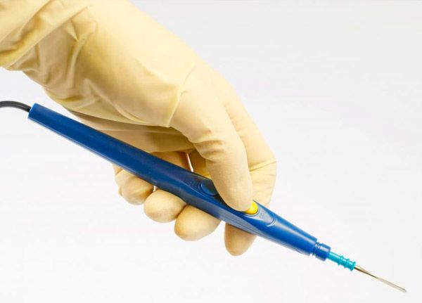
A special medical device, an electrocoagulator, is used for removal. It allows you to control the frequency and strength of the current for performing surgery on different parts of the body. At the end of the device there is a steel loop, which is heated to the required temperature (usually 80 degrees) using electric current and then cauterizes the mole site. The procedure lasts an average of 15-20 minutes. After cauterization, a dense crust forms at the site of mole removal, which is covered with manganese, thereby enhancing crust formation. Under it, there is a process of rapid and even epithelialization. The crust protects the lower layer of the epithelium from infections, so you cannot tear it off prematurely. After 4-5 days, the crust falls off by itself and pink spots remain in their place, which completely disappear within 2 weeks. During the entire period, in order to prevent suppuration, the crust must be lubricated with an antiseptic prescribed by a doctor, for example, a 5% alcohol solution of potassium permanganate.
Contraindications
Like any electrosurgical intervention, mole removal by electrocoagulation has several contraindications. This procedure should not be performed in cases of:
- heart disease;
- herpes of any form in the active stage;
- inflammation in the area of mole formation;
- blood diseases, low blood clotting;
- individual allergy to anesthetics or intolerance to electrical procedures;
- keloid disease;
- acute infectious disease;
The electrocoagulation procedure is strictly prohibited in the case of malignant neoplasms. In such cases, surgical methods of removal are used, which allow cutting out a fairly large area of skin around the tumor to prevent relapse. Electrocoagulation for malignant tumors can provoke the spread of metastases. Also, large moles (from 10 mm) are best removed with a scalpel, and the wounds are sutured with surgical threads to prevent suppuration and rapid healing of the wound. However, after cutting out with a scalpel, a scar remains.
Complications after removal of moles by electrocoagulation
Frequent trauma to a mole, as well as improper removal, can provoke the development of melanoma. This is a very dangerous malignant tumor that degenerates from pigment cells and very quickly metastasizes to other parts of the skin and organs. The insidiousness of melanoma is that externally it can look like a small pigment defect, but internally metastases can already spread to most vital organs. Therefore, if there are any changes in the color, size of the mole or sensitivity of the removal site, you should consult a doctor. High-quality electrocoagulation is considered the best way to prevent melanoma.
Possible complications after the procedure:
- infection - as a result of a poorly performed operation or postoperative care, an infection may be introduced into the wound, which will lead to the development of an inflammatory process. Therefore, do the removal of moles by electrocoagulation only in specialized clinics or professional cosmetology rooms, and strictly follow the doctor's instructions for care.
- scarring - people who are prone to keloid scars are not guaranteed to have their moles removed without a trace. After the procedure, especially if large areas of skin were removed, traces may remain.
Also, the deeper the mole was located in the skin, the more likely it is that a whitish spot will remain at the site of the operation.
The condition of the neoplasms and the site after their removal must be constantly monitored.
Rehabilitation period
During the first few days after the procedure, the place where the mole was cauterized will be painful, reddened and swollen, it is important not to expose it to sunlight or ultraviolet rays, or to wet it. If you are careless and introduce an infection, dangerous suppurations will form under the crust. If the pain does not go away within a few days, or has worsened, you should see a doctor immediately.
If electrocoagulation of a shallow mole is successful, a pale pink spot should remain at the site of the former crust, which will soon disappear and the skin will regain its natural color. High-quality removal of a mole by electrocoagulation and proper postoperative care ensure the absence of complications and scars at the site of the intervention.
Care
After the crust falls off, in order to prevent inflammation, you should not use cosmetic creams, lotions, hard washcloths, scrubs, or visit public baths or saunas for 2 weeks. Also, avoid sunbathing and solariums to avoid pigment spots and relapses. If your doctor instructs you, you can apply sunscreen (at least 60 SPF) or baby cream without additives to the mole removal site before going outside, but do not self-medicate, as this can be dangerous. It is best to agree with the doctor or cosmetologist who performed the operation about his competent monitoring of the wound healing process for two weeks after the operation.
Wounds heal faster in areas where the skin is thinner, so if you have removed moles in different areas at the same time, you should wait with active sports, sun exposure or water treatments until the last wound has healed.
 [ 8 ]
[ 8 ]