Medical expert of the article

New publications
Ultrasound of skin and subcutaneous fatty tissue
Last reviewed: 04.07.2025

All iLive content is medically reviewed or fact checked to ensure as much factual accuracy as possible.
We have strict sourcing guidelines and only link to reputable media sites, academic research institutions and, whenever possible, medically peer reviewed studies. Note that the numbers in parentheses ([1], [2], etc.) are clickable links to these studies.
If you feel that any of our content is inaccurate, out-of-date, or otherwise questionable, please select it and press Ctrl + Enter.
Ultrasound examination of the skin is one of the "classic" diagnostic methods, successfully used for many years in medical and research centers. The principle of ultrasound is the same as in optical coherence tomography, only instead of a light wave, an acoustic wave is used. Ultrasound vibrations during propagation obey the laws of geometric optics. In a homogeneous medium, they propagate rectilinearly and at a constant speed. At the boundary of different media with different acoustic density, some rays are reflected, and some are refracted, continuing rectilinear propagation. The higher the gradient of the difference in acoustic density of the boundary media, the greater the proportion of ultrasonic vibrations is reflected. At the boundary of the transition of ultrasound from air to skin, 99.99% of vibrations are reflected, therefore, before ultrasound scanning, a special gel must be applied to the skin, which plays the role of a transition medium. The reflection of a sound wave depends on the angle of incidence (the greatest reflection will be when the wave falls perpendicularly onto the surface) and the frequency of ultrasonic vibrations (the higher the frequency, the greater the reflection).
Today, ultrasound technologies are actively used to monitor skin edema and wound healing, to study the structure of the skin in diseases such as psoriasis, scleroderma, panniculitis. An important application of the ultrasound method is the detection of tumor formations (melanoma, basal cell carcinoma, squamous cell carcinoma).
Ultrasound examination technique of skin and subcutaneous fat tissue
Skin examination should be performed with high-frequency sensors (15-20 MHz). Ultrasound waves with a frequency of 7.5 to 100 MHz are used for skin examination. The resolution increases with the increase in the frequency of the ultrasound wave, at the same time there is a strong weakening of the echo amplitude in the deeper layers of the skin, so the depth of measurements at high frequency is small.
Skin echo picture is normal
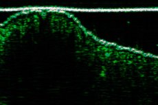
The skin appears as a hyperechoic homogeneous layer.
The thickness of the skin varies depending on the location, it is greater in men than in women.
The subcutaneous fat layer typically appears hypoechoic with alternating hyperechoic fine fibers reflecting connective tissue layers.
 [ 1 ]
[ 1 ]
Pathology of the skin and subcutaneous fat
Edema. With edema, the subcutaneous fat tissue is thickened and its echogenicity is increased.
When edema occurs, connective tissue fibrous bridges appear hypoechoic, while fat layers appear hyperechoic. Edema is usually observed in cellulite, venous insufficiency, and lymphedema.
Foreign bodies. Foreign bodies appear as structures of increased echogenicity surrounded by a hypoechoic rim. The hypoechoic rim that forms around a foreign body is a consequence of an inflammatory reaction.
Wooden and plastic objects appear as hyperechoic structures with a distal acoustic shadow effect.
Metal and glass objects produce a "comet tail" type reverberation effect.
Lipomas. Lipomas may appear as formations in the thickness of the subcutaneous fat. Their echogenicity may range from hyper- to hypoechoic. They may be limited and enclosed in a thin capsule or diffuse without a clear capsule.
Hematomas. Hematomas appear as anechoic or hypoechoic fluid-containing structures. They are formed as a result of trauma. Depending on the duration, the internal structure of hematomas may change.
Nevi. There is a pigmented "head" of the nevus on the skin surface. However, the base of the nevus is located deep in the subcutaneous fat. As a rule, nevi are oval in shape, have clear contours, and are delimited from the surrounding tissues by a thin capsule. Their echogenicity is low. There is a distal effect of echo signal amplification.
Fibromas and fibrolipomas. Fibromas look like hypoechoic oval-shaped formations in the thickness of the subcutaneous fat. As a rule, a capsule is detected that limits the formation. Fibromas have a cartilaginous density by palpation and are limitedly mobile. Sometimes, it is possible to visualize a single vessel on the periphery of the formation.
Ossifications. Hyperechoic inclusions in the thickness of the skin and subcutaneous fat may form after an injury, due to the deposition of calcium salts in the scar, with diffuse systemic skin diseases (scleroderma). Sometimes, they form independently, like sesamoid bones. Often, sesamoid bones are detected in front of the patella.
Angiomas. These are vascular formations consisting of various structural elements (hemangiomas, fibrolipoangiomas, angiomyolipomas, lipangiomas, etc.). The main characteristic is the presence of vessels at the base of the formation.