Medical expert of the article

New publications
Verrucous nevus
Last reviewed: 12.07.2025

All iLive content is medically reviewed or fact checked to ensure as much factual accuracy as possible.
We have strict sourcing guidelines and only link to reputable media sites, academic research institutions and, whenever possible, medically peer reviewed studies. Note that the numbers in parentheses ([1], [2], etc.) are clickable links to these studies.
If you feel that any of our content is inaccurate, out-of-date, or otherwise questionable, please select it and press Ctrl + Enter.
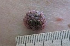
Verrucous epidermal nevus (nevus verrucosus) is a benign, pigmented growth on the skin surface that resembles a wart (in Latin, wart is verruca), and is therefore also called a warty birthmark. Verrucous epidermal nevus (VEN) is a skin disorder that usually occurs at birth and is often resistant to treatment. [ 1 ]
Epidemiology
Verrucous nevi, according to some data, are present in 0.1% of the population; they account for about 6% of all epidermal nevi. [ 2 ]
Causes verrucous nevus
Despite the histological differences between different types of moles on the body – limited areas of abnormal accumulation (hypertrophy) of non-malignant skin cells – the reasons for their appearance are identical.
Nevogenesis, that is, the development of warty nevi, as well as other melanocytic and keratinocytic nevi, is a complex process. Today, it is believed that congenital moles as a skin defect appear sporadically due to errors in the migration of melanocytes (cells that produce skin pigment) from the neural crest during the embryonic period.
More information in the article – Causes of moles.
Risk factors
The main risk factors for the development of congenital nevi are considered to be heredity, pregnancy pathologies and teratogenic effects on the fetus, which negatively affect the entire process of embryonic development.
The risk of developing warty moles is increased by exposure to high doses of ultraviolet radiation and especially sunburn, which activates the function of skin melanocytes and increases the synthesis of melanin.
Pathogenesis
Nevus cells – nevocytes – are a type of melanocyte, but they are larger than typical pigment cells – with more voluminous cytoplasm and large granules, do not have dendrites, deposit melanin and are localized in clusters at the border between the dermis and epidermis and in the dermis.
It is now known that in 40% of cases, the pathogenesis of epidermal nevus – including warty nevus – is associated with genetic mosaicism, splicing or mutations in the FGFR3 and PIK3CA genes. The FGFR3 gene encodes the formation of a protein – fibroblast growth factor receptor type 3 (FGFR-3), which plays an important role in the cellular processes of embryogenesis, as well as in cell proliferation (division), their differentiation and angiogenesis (formation of blood vessels). [ 3 ]
The PIK3CA gene encodes the synthesis of the p110 alpha protein (p110α), a subunit of the phosphatidylinositol 3-kinase enzyme, which ensures the transmission of intracellular chemical signals that regulate cell growth, division, migration, and apoptosis. [ 4 ]
It has been established that mutations associated with nevus affect only the cells of the mole and are not detected in the cells of normal skin.
Symptoms verrucous nevus
Usually, a warty nevus is already present in newborns or begins to appear in infancy, slowly increasing in size later. For adults, the appearance of this type of nevi is uncharacteristic.
The symptoms of such a nevus are hyperpigmented papules of a yellowish-brown color, merging into plaques of various sizes and shapes with a bumpy or granular surface. The formations can be single, but more often they are multiple. Their specificity is a linear or curved-intermittent configuration - along the so-called Blaschko lines (the directions of migration of embryonic cells from the neural crest). [ 5 ]
Warty nevi can be unilateral, bilateral, or located on any area of the skin, such as along an entire limb, on the chest, abdomen, or back.
Complications and consequences
Verrucous epidermal nevi are often resistant to treatment and have a high recurrence rate. [ 6 ] Verrucous nevus is not prone to malignant transformation (i.e. it is not melanoma-dangerous). Negative consequences and complications of this type of epidermal nevi can be a consequence of traumatic impact and infection of the damaged area of skin. Read also: Dangerous and harmless changes in moles, Why does a mole itch and what to do?
Diagnostics verrucous nevus
In addition to a visual examination of the patient’s skin, diagnostics include:
See also the publication – Diagnosis of moles
Differential diagnosis
Differential diagnosis should distinguish warty nevus from congenital linear porokeratosis, Solomon syndrome (Schimmelpenning-Feuerstein-Mims syndrome), actinic keratosis, linear lichen (lichen), ulcerative stage of pigment incontinence, ichthyotic bullosis of Siemens. [ 7 ]
Who to contact?
Treatment verrucous nevus
As with other moles, the treatment for verrucous nevus is its removal, i.e. surgical treatment, more details – Surgical removal of moles. However, surgical removal may not be possible when the skin lesion is very extensive, and this may lead to scarring. Many other treatments have been reported, including topical medications, cryotherapy, [ 8 ] laser treatment, [ 9 ] photodynamic therapy and chemical peels with varying clinical outcomes. [ 10 ], [ 11 ]
Read also: Mole Removal: An Overview of the Main Methods
However, as clinical practice shows, after removal of epidermal nevi, their recurrence is possible.
Forecast
In the presence of such a nevus, the prognosis can be considered favorable, since at a certain stage the formation stops growing, and its transformation into melanoma is practically excluded.