Medical expert of the article

New publications
Causes of moles
Last reviewed: 04.07.2025

All iLive content is medically reviewed or fact checked to ensure as much factual accuracy as possible.
We have strict sourcing guidelines and only link to reputable media sites, academic research institutions and, whenever possible, medically peer reviewed studies. Note that the numbers in parentheses ([1], [2], etc.) are clickable links to these studies.
If you feel that any of our content is inaccurate, out-of-date, or otherwise questionable, please select it and press Ctrl + Enter.
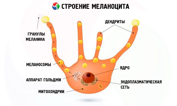
It is generally accepted that the causes of the appearance of moles on the body, which can form on any part of it, are rooted in the benign local proliferation of melanocytes - dendritic cells of the basal layer of the epidermis.
These are the only cells that synthesize the pigment melanin, which protects the skin from ultraviolet rays and determines the color of the skin, hair and eyes.
In terms of structure and properties, melanin is a UV-filtering biopolymer that is obtained through a multi-stage biochemical transformation of the α-amino acid tyrosine; the pigment is deposited in the organelles of melanocytes – melanosomes, and gets into the upper layers of our skin thanks to keratinocytes.
Accumulating in one place, melanocytes form moles, and their average number in one person is from 30 to 40.
Key causes of moles in adults and children
In order to find out the true reason for the appearance of moles, biologists and doctors have conducted and are conducting (and will continue to conduct) numerous biochemical and genetic studies.
At the same time, experts remind us that the skin is a vital multifunctional organ, the formation of which occurs during the process of embryogenesis, that is, during the development of the human embryo.
Most birthmarks appear during the first 20-30 years of a person's life, and, according to statistics, only one in every 100 babies has birthmarks at birth. And the reasons for the appearance of birthmarks in a child, that is, congenital nevi (in Latin, naevus means "birthmark") are associated with a minor defect in embryonic development in the first twelve weeks of pregnancy.
Melanocytes, which produce skin pigment, are formed from melanoblasts, cells of the neural crest, which, in the early stages of embryogenesis, are dispersed along the upper (dorsal) part of the neural crest into various parts of the body (squamous epithelium of the skin and mucous membranes, hair follicles, tissues of the arachnoid membrane of the brain). In the basal layer of the epidermis, melanoblasts mature into melanocytes, which are capable of producing melanin. The defect is believed to lead to accelerated proliferation of melanocytes.
This means that there is an excess of them, and the “excess” melanocytes are not distributed evenly in the skin, but are collected together – in nests, clusters, islands – in the very top layer of the skin and even protrude from it.
Recent studies shed some light on the matter. Some melanocytes arise from melanoblasts that migrate ventrally, along the underside of the neural tube and then along the nerves. These melanocyte precursor cells give rise to the peripheral nervous system and the adrenal medulla. They thus end up in the sheaths of nerves and axons, among the Schwann cells, and are capable of producing melanocytes after birth.
There is scientific evidence that melanocytes in moles are modified into so-called dermal nevus cells. This type of melanocyte differs from the usual one in its size, volume of cytoplasm and absence of processes (dendrites). They are usually located at the border of the transition of the dermis to the epithelial tissue, and depending on the degree of maturity, they can be further classified as epithelioid, lymphocytoid and neuroid. It is claimed that nevus cells are capable of migrating, penetrating into the lymph nodes and even into the thymus gland (thymus), where immunocompetent cells - lymphocytes - are formed and mature.
Today it has been established that in 60% of cases the causes of moles in adults and children are hereditary. More than 125 different genes are already known that regulate pigmentation either directly or indirectly. Many of these genes control the differentiation of melanocytes or affect the biogenesis and function of melanosomes, and also ensure the participation of hormones, growth factors, transmembrane receptors (EphR, EDNRB2, etc.), transcription factors (such as MITF, Sox10, Pax3, etc.) in the biochemical processes of pigmentation and proliferation of epithelial cells. The interaction of all of the above determines the causes of the appearance of new moles.
By the way, about hormones. Hormonal changes during pregnancy and in patients with diabetes often contribute to the formation of moles. And hormonal causes of moles in children and adolescents are explained, first of all, by the activity of hormones and biochemical growth factors (for example, the SCF factor of stem cells): after all, children grow, and the area of the skin is constantly increasing. Also, in a growing organism, melanocortins produced by the pituitary gland are very active - hormones that specifically stimulate the synthesis of melanin (they also affect the production of corticosteroids in the adrenal cortex and the activity of lipid metabolism in the cells of adipose tissue).
Under the influence of solar radiation, melanin synthesis increases (and we see this when a tan appears). All this is the result of the activation of tyrosinase in melanocytes, which provides increased protection of the skin from UV. Some scientists suggest that excessive solar radiation may play a certain role in the formation of acquired moles. So far, the biomechanics of the interaction between genetic structure and general exposure to ultraviolet rays has not been clarified. However, the fact that this is exactly the case is supported by the almost complete absence of moles on the buttocks...
Causes of moles on the neck, face and armpits
Almost everyone is interested in answers to three questions:
- Are there any special reasons for the appearance of moles on the face?
- What are the causes of moles on the neck?
- What are the reasons for the appearance of moles under the armpits - in such an inconvenient place, which, in general, is not exposed to the sun?
We will try to answer them based on what is already known to clinical dermatology regarding the formation of epidermal nevi in the indicated localization.
Melanocytes are located between basal keratinocytes in an approximate ratio of one to ten and distribute melanin through their elongated processes (dendrites), as well as through direct cell contacts. As is known, keratin cells of the skin in the upper layers of the epidermis replace each other quite quickly and, rising upward (to the stratum corneum of the skin) carry away the captured melanin - to form a barrier from ultraviolet rays.
At the same time, in different areas of the epidermis, the content of melanin and the number of cells producing it are different: in the skin of the head (including the face), as well as the neck and hands, there are twice as many melanocytes as in other parts of our body. Obviously, this is due to the fact that these areas are much more often open, and they are exposed to the most sunlight.
Among the unproven versions of the cause of the appearance of moles on the face, there is a suggestion that the process of formation of nevus skin cells is facilitated by increased metabolism in the cells of the epidermis - due to the stressful effects of changes in temperature and air humidity on the skin of the face, as well as constant stretching and compression of the skin by facial muscles.
In addition, there is an opinion that there may be reasons for the appearance of moles on the neck associated with disturbances in the formation and distribution of melanin in areas of the epidermis directly above the nerves of the cervical nerve plexus (see above - about the migration of melanoblasts during embryonic development). These are branches of the motor, cutaneous and phrenic nerves, which are connected by loops and are located on the neck (back, front and on both sides).
But the reasons for the appearance of moles under the armpits, researchers tend to see in the presence of hair follicles and glands in the skin of the armpits - sweat and apocrine. But the specific mechanism for the formation of nevi under the armpits has not yet been studied. Moreover, it remains unknown how the flow of melanocytes into the epidermis is controlled, although, of course, a regulatory scheme for this process exists.
 [ 1 ]
[ 1 ]
Causes of Pink and Red Moles
The most probable reason for the appearance of red moles is that the "body" of the nevus may be not only melanocytes, but also cells of the epidermal connective tissue, adnexal fibers, and vascular elements. The so-called vascular nevi (nevus vascularis) appear as reddish swellings or spots of varying sizes on the skin due to capillary hypertrophy - the proliferation of blood vessels in the skin.
In addition, there may be a connection with a deficiency of blood clotting factors and vitamin K, which leads to increased bleeding when the walls of skin capillaries are damaged, partially entering the formation.
According to dermatologists, red moles are characteristic of diagnoses such as autoimmune rheumatoid arthritis or systemic lupus erythematosus.
The causes of red convex moles are similar. Their “convexity” (as in the case of brown moles) is the result of the melanocytes often being located significantly above the dermoepidermal junction and localized in the upper layer of the epidermis, including the granular zone and stratum corneum.
Also read – Red mole or angioma
The causes of pink and red moles do not exclude the influence of the composition of the melanin produced. Melanin can be either brown-black (eumelanin) or reddish-orange (pheomelanin). In the latter case - especially in redheads and natural blondes - moles are often light beige or pink.
Causes of hanging moles
It is unnecessary to say that the reason for the appearance of a mole on a stalk, as well as the reasons for the appearance of hanging moles on the neck, have been thoroughly studied. Although a lot of attention is paid to the study of the etiology of this type of epidermal nevi.
Thus, an association of melanocytic nevus with eccrine sweat glands has been identified, which is expressed not only in the capture of the gland itself by the body of the mole (which can be located in the center of the mole), but also in the exit of nevus cells in the form of a node to the outside - through the eccrine ducts.
In other cases, the infiltration pattern results in a linear pattern of intradermal nevus cell distribution. Going beyond the dermo-dermal border and the papillary layer of the skin, a group of such cells penetrates the surface, expanding the part of the epidermis between the collagen fibers. Moreover, intradermal nevus cells can form a pigmented dome-shaped or papillomatous papule (up to 1 cm in diameter) equipped with a stalk. A mollusc-like form with a wide base is also possible, with a color from light brown and black to whitish or pink-red.
Hanging moles can form anywhere, but their “favorite places” are the neck area, armpits and skin in the perineal area.
In the middle of the last decade, researchers from King's College in London examined 1,200 non-identical female twins aged 18 to 79 and found that those who had more moles on their bodies also had stronger bones, i.e. they were less likely to develop osteoporosis. In addition, older women with more than 60 moles had less wrinkled skin and looked younger than their years... It turned out that people with a large number of moles have chromosomes with unusually long telomeres - the end sections of DNA polymerase, which prolongs the period of active replication and postpones many age-related processes in the body.
And dermatologists advise – regardless of the time and reason for the appearance of moles – to contact specialists for any changes in epidermal nevi, since the risk of developing skin cancer associated with the presence of moles is quite high.