Medical expert of the article

New publications
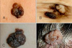
Borderline intradermal nevus.
Last reviewed: 04.07.2025

All iLive content is medically reviewed or fact checked to ensure as much factual accuracy as possible.
We have strict sourcing guidelines and only link to reputable media sites, academic research institutions and, whenever possible, medically peer reviewed studies. Note that the numbers in parentheses ([1], [2], etc.) are clickable links to these studies.
If you feel that any of our content is inaccurate, out-of-date, or otherwise questionable, please select it and press Ctrl + Enter.
Intraepidermal or borderline nevus is one of the many varieties of nevi, which has its own clear characteristics and features. This neoplasm is small, but quite dangerous: it has a tendency to grow and become malignant. Because of this, dermatologists classify borderline nevus as a melanoma-hazardous growth.
Epidemiology
Borderline nevi are common, accounting for approximately 30% of all such growths. They sometimes appear as multiple lesions, but are more commonly found singly. The size of a single growth does not exceed ten millimeters. Epidermal nevi have an incidence of approximately one in 1,000 live births and affect males and females equally. [ 1 ], [ 2 ] It is estimated that one third of people with epidermal nevi have involvement of other organ systems; hence, the condition is considered epidermal nevus syndrome (ENS), and it has been reported that up to 10% of people with epidermal nevi may develop additional features of the syndrome. This syndrome is usually evident at birth (due to skin lesions that are most often seen in the midface from the forehead down to the nasal area) and is often associated with seizures, intellectual disability, eye problems, bone malformations, and brain atrophy. [ 3 ]
The neoplasm can appear at any age, although it is most often detected in patients aged 14-25 years. [ 4 ]
Borderline nevus most often occurs:
- in people who frequently sunbathe, visit solariums, or work outdoors;
- in people who are forced to regularly come into contact with chemical solutions and substances;
- in patients suffering from endocrine diseases or those undergoing treatment with hormonal drugs.
Causes borderline nevus
Scientists are sure that a border nevus is “formed” already during the intrauterine development of the fetus. The cells of the future neoplasm are the precursors of healthy melanocytes, which, however, are retained in the deeper layers of the dermis and are formed in the form of clusters. Under the influence of certain factors, such cells begin to produce pigments, which we notice on the skin as moles.
Sun rays play a significant role in the appearance of border nevi. They can be safely called the main activators of nevus cells that accumulate in the layers of the dermis. With a sufficient dose of solar radiation, these structures begin to accelerate the production of melanin, which is found on the skin as a well-known birthmark.
In addition, altered hormonal activity can also be a stimulating factor. For example, in pregnant women, in teenagers, or during hormonal therapy, the number of nevi on the body increases, and existing border nevi can grow or change their configuration.
Risk factors
Almost all scientists support the theory of the congenital nature of the border nevus. Even despite the fact that the growth can appear ten or twenty years after a person’s birth. The fact that the nevus sooner or later reveals itself may be due to the action of certain factors:
- hormonal changes – for example, with the onset of pregnancy, menopause, lactation period, during hormone therapy, etc.;
- excessive tanning - both under the sun's rays and in a solarium;
- genetic disorders accompanied by abnormal development of melanoblasts;
- dermatitis and other dermatological diseases (acne, eczema, etc.);
- damage and injury to the skin;
- viral infections.
In addition, people who work or have regular contact with chemicals and other toxic substances are at risk.
Pathogenesis
The borderline nevus is initially formed from melanocytes, which begin their development in the prenatal stage. The neoplasm is formed from nerve fibers. Normally, each cellular structure has its own canal for the removal of pigment, but in the altered cells there are no such canals. Therefore, melanin does not come out, but accumulates in a limited area, which explains the formation of dark spots. Genetic and clinical mosaicism has been described. [ 5 ] It has been found that germline mutations in the FGFR3 gene are the etiology of congenital epidermal nevus. [ 6 ]
A border nevus is formed at the borders of the upper and middle skin layers, bypassing the basal layer. Most often, they talk about the congenital nature of the growth, although it can appear in teenagers and even in 20 or 30 years.
In terms of the degree of danger of malignant transformation, borderline nevus is placed on the same level as nevus of Ota, Dubreuil's melanosis, and giant pigmented nevus. [ 7 ]
Symptoms borderline nevus
The most common site of involvement was the head and neck, and 13% of patients had widespread lesions. [ 8 ] A junctional nevus appears as a flat, nodular formation with a gray, black, brownish tint. The size of the nevus varies from a couple of millimeters to one centimeter, although some experts also talk about larger spots.
The growth is smooth, dry, and sometimes slightly uneven on top. The main distinguishing feature: hair never grows on a border nevus, although the growth can be located almost anywhere on the body, even on the feet or palms.
The neoplasm is usually single, but multiple locations also occur.
The first signs of degeneration of a border nevus are a change in color and/or its size, the formation of cracks, ulcers, tubercles on its surface, the appearance of redness, and the loss of clear contours. These symptoms indicate the need to urgently visit a dermatologist.
Stages
The transformation of a borderline nevus into a malignant tumor usually occurs through several stages:
- Initial stage of development, without metastases. The duration of the stage varies from 12 months to five years. The chances of cure are up to 99%.
- The nevus becomes convex to about 4 mm. Malignant transformation into a dysplastic process is observed within several months. The chances of cure are up to 80%.
- Within 1-3 months, metastases begin to spread, which are found in the lymphatic system, brain, internal organs. The nevus itself ulcerates. The chances of recovery are no more than 50%.
- The aggressive stage, which ends within a few weeks, results in the death of the patient in 85% of cases.
Forms
Experts distinguish between potentially dangerous and safe borderline nevi, according to the degree of probability of their transformation into malignant melanoma. In addition, other types of neoplasms are also distinguished. [ 9 ]
- An acquired borderline nevus is a neoplasm that was discovered not at birth, but somewhat later - for example, after a couple of years, or even in adulthood. However, doctors say that this does not mean that the nevus was not formed in utero. It is just that a combination of factors contributed to the later manifestation of the growth.
- A borderline pigmented nevus is a pigmented nodular formation, up to 10 mm in size, with any location on the body. A type of such a neoplasm is considered to be a cocarded nevus - a growth with increased pigmentation along the peripheral border, which gives it a ring-shaped appearance. Both pigmented and cocarded nevi are melanoma-hazardous elements.
- Melanocytic junctional nevus is a neoplasm caused by excessive proliferation of epidermal melanoblasts, which in turn is caused by a failure of gene regulation. Initially, a junctional nevus is formed in the epidermis. Over time, some of the melanocytes are transported to the dermis, and another part remains in the epidermal layer: this is how a complex melanocytic nevus is formed. [ 10 ], [ 11 ]
- Nevus with borderline activity is characterized by predominant intradermal structures. In this case, borderline activity refers to the proliferation of melanocytes, which can be focal or widespread.
- A borderline dysplastic nevus is a pigmented mole of borderline location, irregular ovoid shape, with unclear contours and uneven pigmentation (the central part is one color, and the edges are another color). Such a neoplasm is often classified as a clinical marker of increased risk of melanoma development. [ 12 ]
Complications and consequences
The most undesirable and unfavorable complication of a borderline nevus is its transformation into a malignant tumor – melanoma. Such a transformation does not happen “out of the blue”: it requires the influence of certain factors that create the necessary conditions for degeneration. For example, the risk of malignancy increases significantly if the nevus is regularly exposed to sunburn or trauma. [ 13 ]
To avoid complications, doctors recommend removing border nevi, even if they do not bother or change. Transformation into melanoma, melanoblastoma, skin cancer is difficult to treat and often leads to the death of the patient. People with fair skin, light or red hair, as well as those who have a large number of different moles on their body, including border nevi, should be especially careful.
Recurrence of borderline nevus
In about 80% of patients, a border nevus may recur after its removal by laser or destructive means. The growth develops in the same or another place. Some patients have to get rid of the obsessive nevus several times.
Doctors note: the most radical method of removal is the surgical method, when the neoplasm is excised together with the surrounding healthy tissue, the volume of which depends on the shape of the nevus. The larger the growth, the more it is prone to recurrence. If a person has already had relapses, then he should pay special attention to the prevention of complications:
- spend less time in the sun, especially during active hours (from 11:00 to 16:00);
- eat quality food rich in vitamins and minerals;
- give up bad habits, lead a healthy lifestyle;
- try to wear high-quality natural clothing, do not injure your skin, even if there are no nevi or birthmarks on it.
Diagnostics borderline nevus
Diagnosis of a borderline nevus begins with anamnesis, external examination and dermatoscopy. Histology is performed only after removal of the neoplasm, but not earlier than this moment. Histopathological changes associated with the aging of melanocytic nevi, such as fatty degeneration, fibrosis and neural changes, are detected in the lobular intradermal nevus. [ 14 ] The fact is that the process of taking material (biopsy) is also a damaging factor that can cause subsequent malignant transformation of the growth. [ 15 ]
Blood tests include the following research options:
- blood test for coagulation quality;
- blood for tumor markers;
- blood test for LDH (lactate dehydrogenase).
Instrumental diagnostics primarily involves dermatoscopy, a method that helps examine changes within the skin that are not visible to the naked eye. Additionally, the doctor may prescribe an ultrasound examination of the nearest lymph nodes, chest X-ray, osteosyntigraphy, to rule out malignant processes in the body.
Differential diagnosis
Differential diagnostics should be carried out with other forms of hyperpigmentation - and first of all, with chloasma, which looks like a mole, or with hemangioma. But it is more important to pay timely attention to the degeneration of a borderline nevus into malignant melanoma. The tumor process sometimes develops almost imperceptibly, against the background of a minor dysplastic syndrome: the contours of the spot expand slightly, the surface becomes bumpy, the healthy skin located nearby turns red. Since degeneration often occurs after mechanical trauma to the skin, it is important to regularly examine the growths that form on the plantar and palmar surfaces of the extremities, between the fingers and near the nail plates. In such places, it is recommended to remove moles, regardless of their type and degree of danger.
Who to contact?
Treatment borderline nevus
After conducting the diagnosis, the doctor will consider all possible treatment options, although a conservative method is usually not discussed: the border nevus is removed in one of the following ways:
- Cryodestruction is a procedure of freezing the growth using liquid nitrogen (less often using carbonic acid or ice). [ 16 ]
- Electrocoagulation is a method that involves the destruction of a neoplasm using high temperature, caused by the action of a directed current. [ 17 ]
- Laser removal is one of the most popular methods, which involves “evaporating” the affected tissue with a directed laser beam.
- Radiosurgical procedure – involves excision of the growth with a certain length of radio waves, using the Surgitron hardware device.
Medications can be recommended only at the recovery stage after removal of the border nevus.
Physiotherapeutic treatment involves the following procedures:
- UHF coagulation – involves the use of an electrode with a high-frequency current supply of 27.12 MHz and 1 mA. Upon completion of the procedure, the coagulation area is treated with a 5% solution of potassium permanganate. [ 18 ]
- Laser thermocoagulation is performed using continuous and pulsed optical irradiation of the infrared range, with a maximum power of 3-5 W and a focused beam diameter of 0.25-0.5 mm, with an emitted power of 10-15 W. [ 19 ], [ 20 ], [ 21 ]
Medicines that a doctor may prescribe
To speed up the healing process after removal of a border nevus, the doctor may recommend the use of the following medications:
- vitamin supplements to improve plastic metabolism (folic acid, B vitamins, ascorbic acid, tocopherol);
- non-steroidal anabolic agents (Riboxin, potassium orotate, methyluracil);
- biogenic stimulants (aloe extract, FiBS, Plasmol);
- immunomodulatory agents (Timulin, Pyrogenal, Levamisole);
- nonspecific regenerating agents (sea buckthorn oil, Apilak, Rumalon, Actovegin).
Examples of the use of these medications are highlighted in the following table:
Methyluracil |
Adult patients are prescribed one tablet 4 times a day for a month. The treatment may be accompanied by headaches, heartburn, allergic reactions. |
Aloe extract |
Administer subcutaneously daily 1 ml, for several weeks. Possible side effects: dyspepsia, changes in blood pressure, allergy, dizziness, itching. |
Timalin |
Administered intramuscularly with saline, 5-20 mg daily. The course of treatment is from three to ten days. Side effects may be limited to a local reaction in the injection area. |
Actovegin |
Take 1-2 tablets three times a day for 4-6 weeks. The medicine is well tolerated, allergies and fever rarely occur. |
Vitamin E |
The dose of the drug is selected individually, not exceeding the daily amount of 1000 mg. Possible side effects include nausea, headache, fatigue, and allergies. |
Folk remedies
There are many folk recipes that involve the impact on birthmarks and nevi. Doctors do not approve of most of them - especially when it comes to melanoma-hazardous neoplasms, which include borderline nevus. In relation to them, it is better to use radical removal, seeking help from a surgeon.
However, many patients try to get rid of moles in the following ways:
- Mix equal parts of linseed oil and flower honey. Rub the mixture into the nevus area three times a day, every day.
- Wipe the growth with fresh pineapple juice several times a day.
- Every day, drip a drop of onion juice or apple cider vinegar onto the nevus.
- Lubricate the mole with lemon juice and garlic.
- Grind 100 g of cherry pits into powder, pour in 500 ml of any vegetable oil, keep in the refrigerator for a couple of weeks. The resulting oil is used daily for application to nevi: leave on the growth for about twenty minutes, then wash off with water.
You should not rely on folk methods if the borderline nevus has begun to show at least one sign of malignant degeneration - for example, it has begun to increase in size, changed shape or color, has become blurry, swollen, etc. It is always better and safer to consult a doctor in advance.
Surgical treatment
The treatment of choice for small epidermal nevi is surgical excision.
Surgical excision, dermabrasion, cryosurgery, electrosurgery, and laser surgery have been used to treat epidermal nevi.[ 22 ],[ 23 ],[ 24 ] Dermabrasion, if superficial, is associated with a high recurrence rate, and deep dermabrasion may result in thickened scars. Cryosurgery has similar limitations with risks including slow healing, infection, swelling, and usually abnormal skin coloration. Physicians have been performing laser treatments for epidermal nevi for decades. Recent advances in laser technology have improved the ease, precision, and safety of such procedures. Several reliable and effective treatments for epidermal nevi have been developed using CO2, long-pulsed Nd:YAG, and 585 nm pulsed dye lasers. However, recurrence may occur months to years after epidermal nevi are removed by any method. [ 25 ], [ 26 ], [ 27 ], [ 28 ]
Surgery is an old and most effective way to get rid of various types of moles and warts, including border nevus. Preparation for the procedure is simple and short. The skin is treated with a special antiseptic, and local anesthesia is performed. When the anesthesia takes effect, the surgeon excises the nevus with a scalpel, capturing a little healthy surrounding tissue - for a more complete and 100% removal of the growth.
Surgical treatment has its advantages:
- recurrence of borderline nevus is excluded;
- the neoplasm can be sent for histology;
- the intervention is performed in an outpatient setting; there is no need to go to hospital.
The operation is not without its drawbacks, for example:
- the suture takes a little longer to heal than with other removal methods – approximately one month;
- If not properly cared for, there is a risk of suppuration;
- the formation of an unaesthetic scar is possible.
However, with large nevi, doctors insist on surgical intervention. This is the most reliable way to get rid of the problem forever, prevent malignancy and relapse of the neoplasm.
Prevention
It is almost impossible to prevent the formation of a border nevus. However, patients prone to the appearance of moles should be vigilant and carefully examine their body for changes and malignant transformation of pigmented neoplasms.
For preventive purposes, it is necessary to adhere to the following recommendations:
- avoid damaging the skin, and in particular any nevi;
- avoid prolonged exposure to sunlight, do not go to a solarium, do not allow sunburn;
- When working with chemicals and toxic substances, wear protective gloves;
- harden yourself, strengthen your immune system, eat well and nutritiously.
If the border nevus is damaged for any reason, you should seek medical help from a dermatologist or oncologist. He will examine the growth and decide whether it needs to be removed.
Forecast
Doctors advise not to forget that a borderline nevus can degenerate into a malignant neoplasm, regardless of age. Therefore, you should always be attentive and have moles and spots examined by a dermatologist or oncologist at least 1-2 times a year. If suspicious symptoms are detected, it is better to remove the growth without waiting for further unfavorable development of the process.
A borderline nevus is a melanoma-dangerous pathology. But this does not mean that the transformation will necessarily occur: most patients live with such formations, and sometimes do not even suspect their potential danger. Therefore, there is no need to panic. The main thing is to regularly examine the skin, pay attention to all existing nevi, and record any changes on their part.