Medical expert of the article

New publications
Fibroepithelial nevus
Last reviewed: 12.07.2025

All iLive content is medically reviewed or fact checked to ensure as much factual accuracy as possible.
We have strict sourcing guidelines and only link to reputable media sites, academic research institutions and, whenever possible, medically peer reviewed studies. Note that the numbers in parentheses ([1], [2], etc.) are clickable links to these studies.
If you feel that any of our content is inaccurate, out-of-date, or otherwise questionable, please select it and press Ctrl + Enter.
Among the large number of various hypertrophic changes in the skin, dermatologists distinguish fibroepithelial nevus – a common type of pigmented convex moles.
Epidemiology
According to some data, fibroepithelial nevi appear twice as often in women as in men; their peak development occurs between the ages of 40 and 60. [ 1 ]
In no less than 20% of cases of removal of such moles, their recurrence is observed.
Causes fibroepithelial nevus
Fibroepithelial nevi are formed on the surface of the skin (epidermis) sporadically and are classified as benign moles, which experts consider to be non-melanoma-hazardous formations, that is, they do not lead to the development of skin cancer - melanoma.
When listing the probable causes of the appearance of moles, dermatologists name both developmental anomalies (often caused by genetic factors) and hormonal changes, as well as exogenous risk factors for the appearance of pigmented fibroepithelial nevi on the skin.
These factors include ultraviolet radiation (as is known, excessive UV radiation has a mutagenic effect on the skin), although research data regarding the dose-dependent effect of sun exposure on the appearance of nevi are contradictory. [ 2 ]
In addition, risk factors include ionizing radiation (or increased background radiation) and skin injuries (primarily chemical), seborrheic keratosis. [ 3 ]
Of the most scientifically substantiated versions, only deviations in the development of the skin during the embryonic period can be identified, which, in turn, does not exclude their connection with certain pathologies of pregnancy or teratogenic effects. [ 4 ]
Pathogenesis
Researchers have not fully understood the mechanism of development of nevi, including fibroepithelial ones, but there is no doubt that it is associated with the embryogenesis of the skin.
This is why some experts classify these nodular formations as tissue defects – hamartomas.
In particular, pathogenesis is considered to be the result of certain deviations in the migration of the original cells of the neural crest of the embryo to ectodermal areas (primarily to the skin and central nervous system) and their transformation into melanocytes of the skin, which are special cells of the basal stratum that produce pigments (dark eumelanin and light pheomelanin) that protect the skin of the body from UV radiation.
Probably, during the process of intrauterine histogenesis, for some reason, connective tissue cells that make up the fibrous fibers of the underlying dermis (the lowest layer of the skin) can penetrate the epidermis through the basal membrane. Or, which also fits in well with this version, the protoplasmic processes of the basal membrane, which are formed in the skin structure in the early period of intrauterine development and have reticular fibers, locally change direction - towards the epidermis.
In cases of acquired nevi, an unknown signal is thought to trigger melanocyte proliferation.[ 5 ]
Symptoms fibroepithelial nevus
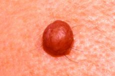
It is difficult to identify the symptoms of asymptomatic convex fibroepithelial nevi present on the skin. Such moles are divided into congenital and acquired, but in both cases these formations on the skin of the body or face have a clearly defined round shape and a wide base (pedicle); the maximum diameter is 10-12 mm; their surface is most often smooth (with a normal skin pattern), but can also be bumpy.
Many moles have hairs growing from their bodies. These nevi are elastic to the touch, and their color ranges from flesh-colored to pinkish and all shades of brown. These nevi do not cause any sensations. [ 6 ]
Complications and consequences
As already noted, fibroepithelial nevus does not transform into melanoma, although it can be damaged, which causes bleeding and does not exclude the development of inflammation.
But after its removal, complications are possible, more details in the material - Consequences of removing a mole.
Diagnostics fibroepithelial nevus
First of all, dermatologists examine the nevus and perform a dermatoscopy. [ 7 ] All the details are in the publication Diagnosis of moles.
Differential diagnosis
Differential diagnostics should be carried out with such skin formations as dermatofibroma or basalioma, as well as with other types of moles on the body.
Who to contact?
Treatment fibroepithelial nevus
There is no drug treatment for nevi, and surgical treatment involves their removal (usually for cosmetic reasons) using electrocoagulation, laser, radio waves, or scalpel excision. All the necessary information is in the material - Removing moles: an overview of the main methods.
But only a regular surgical excision allows for a histological examination of the mole after its removal and to verify the benign nature of the nevus.
Prevention
There are currently no specific measures to prevent the occurrence of fibroepithelial and other nevi.