Medical expert of the article

New publications
Benign moles
Last reviewed: 04.07.2025

All iLive content is medically reviewed or fact checked to ensure as much factual accuracy as possible.
We have strict sourcing guidelines and only link to reputable media sites, academic research institutions and, whenever possible, medically peer reviewed studies. Note that the numbers in parentheses ([1], [2], etc.) are clickable links to these studies.
If you feel that any of our content is inaccurate, out-of-date, or otherwise questionable, please select it and press Ctrl + Enter.
Causes benign mole
Nevi or moles can appear for various reasons. There are several main factors that can lead to the appearance of benign formations. These include:
- developmental defects;
- genetic predisposition;
- negative impact of ultraviolet radiation;
- mechanical and chemical injuries;
- hormonal imbalance;
- viral and bacterial skin lesions.
The main reasons for the appearance of benign moles are developmental defects. In the human body, cell division may fail, resulting in the formation of multiple nevi on the body. They can be both congenital and acquired. In 60% of cases, problems with cell division lead to the appearance of moles.
Genetic predisposition. Nevi are inherited. If any family member has moles, the likelihood of multiple moles appearing in a newborn increases. In many cases, the formations are benign.
Negative impact of ultraviolet rays. Ultraviolet radiation stimulates the work of melanocytes. They are responsible for the production of pigmentation and changes in the shade of the skin. Increased exposure to ultraviolet radiation can provoke the production of melanotropic hormone, which will lead to the formation of nevi.
Mechanical and chemical damage to the skin plays an important role in the formation of formations. The damage affects certain layers of the skin, causing an inflammatory process. Intensive production of active substances leads to stimulation of cell growth.
Hormonal imbalance. Hormones often lead to the development of moles. This is observed during puberty and menopause in women. Hormonal changes in the body affect the functioning of the pituitary gland. As a result, neoplasms develop.
Viral and bacterial lesions of the skin. The appearance of moles due to the negative impact of pathogens on the body is extremely rare. The mechanism of mole development is similar to that of injuries.
 [ 3 ]
[ 3 ]
Pathogenesis
A mole is a growth of skin under the influence of certain factors. Visually, the formation looks like a growth on the skin, characterized by compaction. Pathogenesis is the mechanism of neoplasm development, in this case the process is caused by the production of a significant amount of melanin.
Melanin is produced by melanocyte cells. This occurs under the influence of certain factors, most often ultraviolet radiation. Melanotropic hormone, which is actively produced by the pituitary gland, can participate in the process.
Several systems are involved in the process of nevi formation. As for the development of moles, uncontrolled cell division plays a significant role in this process.
Symptoms benign mole
External signs of benign moles or what do they look like?
It is not difficult to notice a mole on the body. Many owners of these beautiful "growths" know about their existence. However, what does a benign mole look like? The formation of large nevi makes it difficult to determine the type of neoplasm. Even experienced specialists are not always able to cope with this task. Some nevi do not have specific signs.
Typically, these are raised lesions on the skin, characterized by a rough or smooth surface. Pigmentation may be pronounced or absent altogether.
How to determine which moles are benign?
Today, there are several types of skin lesions. The characteristics of the moles presented below will help you find out which of them are benign.
- pigmented moles are small, colored. They are located in the thickness of the skin, rarely protruding above the surface;
- papillomas are characterized by roughness with numerous irregularities;
- halo nevus. Characterized by the presence of a depigmented crown located around the mole;
- Mongolian spot. Increased pigmentation is observed in children. In adults, the mole is barely noticeable. The formation is characterized by large sizes;
- fibroepithelial formation. The mole is smooth, not always colored. Its color can vary from pink to light brown;
- blue mole. It is distinguished by its bluish tint and small size;
- pigmented nevus. The appearance depends on the cause of its appearance. In many cases, the mole has a concentrated brown tint;
- giant formation. The mole reaches large sizes, and increased hair growth is observed on its surface;
- Nevus of Ota. Most often formed in the eye socket and jaw area. Pigmented neoplasms are recorded in the eye, nose and oral cavity;
- dysplastic formation. Characterized by irregular shape and uneven coloring.
[ 7 ]
Complications and consequences
Benign moles, if certain rules are followed, rarely become malignant. Negative consequences may arise due to constant mechanical impact on the nevus. Melanoma or malignant mole is formed under the pressure of certain factors.
Cancer is caused by excessive production of melanocyte cells. This process can be influenced by genetic predisposition, the presence of dysplastic formations and the negative impact of sunlight. People at risk should be careful when on the beach and avoid injuries.
Melanoma in its advanced stage is dangerous to human life. It can metastasize and lead to death. In the early stages, the malignant neoplasm is removed.
Is it possible for complications to develop?
The only serious consequence is the transition of a benign neoplasm into a malignant form. Melanoma is the most common complication. Previously, 95% of cases died from the formation. This is due to the danger of a cancerous tumor and people's inattention.
If there are formations on the skin and they are constantly traumatized, it is necessary to monitor the changes. A change in shade, appearance, surface and size is a serious reason to see a doctor.
Benign moles do not carry any other complications.
[ 8 ]
Diagnostics benign mole
To make a diagnosis, it is necessary to conduct a series of diagnostic studies. Without special diagnostics, no doctor will be able to identify the nature of a benign mole. For this purpose, various measures are carried out, including:
- patient interview (collection of a detailed anamnesis);
- examination of a person with a visual assessment of the situation;
- dermatoscopy. Examination of the formation under a microscope;
- phosphorus isotope indication;
- ultrasound;
- X-ray examination;
- determination of body temperature;
- examination of tissue from the affected area.
Before conducting a full examination, the specialist must listen to the patient's complaints and conduct an examination. A detailed description of the diagnostic measures will be presented below.
How to independently identify a benign mole?
A benign neoplasm is characterized by a regular shape. The color of the mole is brown, while its shape is ideal. When mentally dividing the formation into two parts, they should be ideal. How can you independently determine whether a mole is benign or not?
The size of the nevus should not exceed one centimeter. Formations larger than 1 cm are subject to malignancy, which will eventually lead to a transition to a malignant form. The edges of the mole are ideal, there are no sharp edges or roughness. The nevus does not itch or bleed. Hair does not fall out at the site of its formation. If a strange clinical picture appears, there is a high probability of the mole degenerating into a malignant form.
Is it necessary to take tests?
To get a complete picture of the study, the doctor may refer the patient for tests. It is necessary to give blood and urine. The latter type of study is prescribed extremely rarely. This is due to the absence of changes in this pathology.
Tests are often prescribed before surgery or an upcoming biopsy. In this case, obtaining information is aimed at determining the general condition of the body. Often, additional research methods allow us to identify chronic diseases that can make adjustments to the process of nevus formation.
If a mole has developed against the background of a long-term course of the disease, repeated tests are carried out. Bacteriological studies are used as an addition. They allow choosing the correct tactics for treating the underlying disease.
Research through instrumental diagnostics
Instrumental examinations include dermatoscopy, phosphorus isotope indication, echography, X-ray examination, body temperature determination and examination of the affected area (biopsy). Together, the methods allow identifying the nature of the formation and, if necessary, selecting the optimal treatment regimen. Instrumental diagnostics is the most important part of the study.
- Dermatoscopy. The procedure is a thorough examination of the affected area. A special magnifying device is used during the examination. Thanks to it, changes on the surface of the mole can be detected. This method is considered the most effective and painless. It lasts no more than 20 minutes and allows you to get complete information about the neoplasm. There are no contraindications for use.
- Phosphorus isotope indication. The method is highly sensitive, based on the accumulation of radioactive phosphorus by a cancerous formation. The component takes part in the process of cell division. In the presence of a malignant neoplasm, everything happens quickly. Based on accelerated cell division, the specialist makes a preliminary diagnosis - malignant neoplasm.
- Echography. The main objective of the procedure is to determine the exact size of the mole. Echography is used exclusively in the presence of large formations. The accuracy of the study is low, especially in the case of a small nevus. The device cannot detect a flat mole. Echography is used as an auxiliary method, in rare cases.
- X-ray examination. During the examination, the doctor takes a picture using X-rays. The procedure is carried out with high accuracy, which allows you to get photos in different projections. The method has no contraindications. It is used to diagnose metastases in organs with melanoma.
- Measuring body temperature. This is not an ordinary procedure. Skin temperature is measured using a special preparation. In the presence of a malignant tumor, a slight increase is always recorded. This is due to rapid cell division and active metabolism. The technique is painless, fast and accurate.
- Study of the affected area (biopsy). It is carried out when the first signs of degeneration of a mole into a malignant neoplasm appear. Thanks to the study, it is possible to obtain a 100% result. A certain area of tissue is subject to study, which is preliminarily stained and examined under a microscope.
Differential diagnosis
Before sending a person for a detailed instrumental examination, a number of mandatory procedures are carried out. Differential diagnostics includes examining the patient, collecting anamnesis and evaluating visual data.
Data collection or anamnesis. The doctor asks the patient standard questions regarding his neoplasm. Any diagnostic begins with collecting anamnesis. Many answers will allow the specialist to build a certain picture of the person's condition.
Examination of the patient. After collecting the information, the doctor begins the examination. Each type of nevi has its own visual signs. Detailed information was provided above. Based on this data, the doctor makes a preliminary diagnosis for the patient. To confirm or refute it, it is necessary to visit a dermatologist, oncologist, urologist and gynecologist. At the same time, the person is sent for instrumental diagnostics.
The specialist evaluates the detected moles according to several criteria: quantity, consistency, location, size and surface of the formation.
Is it possible to independently distinguish a malignant mole from a benign one and how to do it?
There is a certain algorithm of actions. Thanks to it, it is easy to distinguish a malignant mole from a benign one, both for an adult and a child.
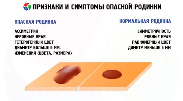
As has been mentioned many times, a common nevus is distinguished by a brown or black shade. The shape of the mole is symmetrical, without any particular protrusions. A benign formation is characterized by clear boundaries. Suspicious moles are always blurred, excessively convex and rough. Their shade can be either brown or red.
The main sign of a safe mole is its uniformity. The size of the formation does not exceed 6 mm. If different shades are observed within one formation, there is a high probability of its degeneration into a malignant formation. An important condition for a benign mole is the absence of discomfort. The nevus should not itch, scratch or cause discomfort. If this clinical picture appears, it is advisable to visit a dermatologist.
Safe moles must be properly looked after. Avoid direct sunlight, mechanical and chemical injuries. Otherwise, the formation may develop into a malignant form.
Who to contact?
Treatment benign mole
Nevi that are subject to malignant transformation must be removed. Removal of benign moles is appropriate in case of high risk of melanoma development. They are subject to surgical excision.
The formation is removed in two cases: if there is a risk of its degeneration into melanoma, and if there is aesthetic discomfort. The method itself depends on the reason for the removal. The specialist decides independently how he will remove the nevus.
For cosmetic indications, the following methods are widely used:
- surgical method;
- cryodestruction;
- electrocoagulation;
- laser removal;
- radiosurgery.
The surgical technique is traditional. It is used to remove a deep or large mole. The main disadvantage of the technique is the presence of traces after the operation. During the surgical intervention, not only the nevus itself is removed, but also part of the surrounding skin.
Cryodestruction is based on the destruction of the formation by liquid nitrogen. Under its influence, the mole gradually wrinkles, forming a dry scab. Over time, healthy tissue grows on it. The procedure is painless and effective.
Electrocoagulation is based on thermal action on the mole. After the procedure, the formation is sent for examination. Gradually, the wound heals and a barely noticeable scar appears in its place.
The best method is considered to be laser removal. It is often used to remove nevi in the face and on open areas of the body. The main advantage of the procedure is the small diameter of the impact and the preservation of surrounding tissues. After removal, nothing remains on the skin.
Radiosurgery. A contactless method of removal performed using a radio knife. Today it is especially popular and is used to remove benign and malignant tumors.
In the case of a cancerous tumor, only surgical removal is used. Excision is performed within the healthy tissue, after which the mole is sent for histological examination.
Removing moles with medication
Drug therapy for the removal of moles is not carried out. For cosmetic purposes and at a high risk of oncology, the nevus is removed by the above-described methods. Medicines are used exclusively to remove papillomas or warts. For this purpose, such drugs as Ferezol, Viferon, Isoprinosine, Panovir and Crinopharm are used.
- Ferezol. Apply to the affected area once. If the lesion is extensive, multiple uses are possible. Before applying, the wart must be steamed. In order to protect the skin around the formation, it is necessary to treat it with zinc ointment or petroleum jelly. There are no contraindications or side effects, the only limitation is that Ferezol is not used to remove warts in children.
- Viferon. It has a pronounced effect, but it will not remove the wart immediately. The cream should be applied for 5-20 days, several times a day. There are no contraindications or side effects. The ointment can be used during pregnancy and breastfeeding.
- Isoprinosine. These are tablets that must be taken orally. The duration of treatment is 14 days, and the drug must be taken 3 times a day, 2 pieces. The visible result will be noticeable in a week. The drug has a pronounced effect on immunity and helps eliminate viruses. For use by children and women during pregnancy, a doctor's consultation is required.
- Panovir. This is a gel with antiviral properties. To remove a wart, it must be applied for 5-10 days, at least 5 times a day. The gel penetrates deeply under the skin and removes the formation along with the root. Before using during pregnancy, a specialist consultation is required.
- Krinofarma. This remedy is the safest. You can use it yourself. The drug freezes the wart, it disappears within 10 days. If the formation is old, the course can be repeated. There are no side effects, the only contraindication is childhood under 2 years.
Traditional methods of treatment
You can remove hated moles without surgical excision and medications. Traditional methods of treatment will allow you to do this without difficulty. However, before using any method, it is advisable to make sure that the formation is not malignant. Otherwise, the risk of worsening the situation increases.
- Lapis pencil. This is the second name for silver nitrate. This substance has been particularly popular for several years. It is used to cauterize moles until the formation begins to decrease. A high concentration of silver nitrate is used for 2-3 weeks. Cauterization is performed 1-2 times a day. If there are no visible improvements, the method should be abandoned.
- Vinegar essence. Treating a mole with vinegar is an effective way to combat it. After a week of use, nevi disappear. However, cauterization is painful, so it is not recommended for children. Vinegar can only be applied once a day. A side effect of the procedure is skin defects.
- Lemon juice. The acid cauterizes the mole, thereby causing tissue destruction. It is necessary to lubricate the formation 4-5 times a day. The removal procedure is long, at least 3 weeks.
Traditional methods do not always help, a positive effect is observed only in 10-15% of all cases. It is advisable to remove nevi using traditional methods. This will reduce the risk of unwanted damage and infection.
[ 11 ], [ 12 ], [ 13 ], [ 14 ]
Treatment of moles with herbs
Traditional methods are based on the use of plants and their juice. Herbal treatment allows you to quickly and painlessly get rid of a nevus. The best methods of removal are considered to be celandine juice and hemp oil.
Celandine juice. This plant has good disinfectant properties. It cauterizes the mole and allows you to get rid of many types of nevi. You can use the juice several times a day, applying it in a thin layer to the surface of the formation. The plant does not have sufficient viscosity, so it quickly rubs off. For durability, the juice is mixed with Vaseline. The resulting mixture is characterized by a pronounced effect.
The second, most common method of removing moles is the use of hemp oil. Due to its properties, the product evaporates the formation. It completely disappears after 4 days or several weeks, depending on the size of the mole.
Prevention
It is impossible to prevent the development of moles. However, with the help of prevention, it is possible to reduce the likelihood of a benign formation degenerating into a malignant one. Recently, a significant increase in the incidence of skin melanoma has been recorded. Women at a young age are susceptible to this influence.
You can prevent melanoma yourself. To do this, you need to limit the time spent in the sun, especially for people with a significant number of moles on the body. You should apply special creams and lotions that reduce the negative impact of ultraviolet radiation. Existing moles must be carefully monitored. It is advisable to avoid constant mechanical and chemical injuries.
If the external indicators of a mole change, you should consult a dermatologist and oncologist. Compliance with all preventive measures will help avoid the development of melanoma.
Forecast
Proper monitoring of the condition of moles will allow the patient to avoid serious complications. The most unfavorable prognosis is with dysplastic formation. Especially if it is not congenital, but acquired. The probability of degeneration of a mole into a malignant formation is 95%.
Most formations are characterized by a favorable prognosis. With constant unfavorable impact on a mole, it can develop into a malignant stage. In this case, it must be removed surgically.
Benign moles are present in every person. The right preventive measures will make a nevus a small highlight, and not a real problem.