Medical expert of the article

New publications
SIascopy of the skin
Last reviewed: 06.07.2025

All iLive content is medically reviewed or fact checked to ensure as much factual accuracy as possible.
We have strict sourcing guidelines and only link to reputable media sites, academic research institutions and, whenever possible, medically peer reviewed studies. Note that the numbers in parentheses ([1], [2], etc.) are clickable links to these studies.
If you feel that any of our content is inaccurate, out-of-date, or otherwise questionable, please select it and press Ctrl + Enter.
Today, modern dermatology offers such a modern method of early melanoma diagnostics as siascopy. Almost any suspicious elements on the skin can be scanned using a siascope. This device will help to reliably determine their structure, assess the likelihood of developing oncopathology and establish the most optimal therapy.
Siascopy is a spectrophotometric intracutaneous analysis that involves the study of pigmented skin elements. The method is based on the interaction of light waves of different lengths with the pigment melanin, hemoglobin and collagen up to two millimeters deep. Siascopy does not cause any discomfort to the patient, as the procedure is quick and painless. [ 1 ]
Indications for the procedure
Siascopy allows visualization of:
- dermatoscopic picture of the skin element with determination of the true color shade and external structure of the neoplasm;
- changes in melanin concentration;
- areas with absence of melanin in the papillary dermal layer;
- zones with higher or lower concentrations of hemoglobin;
- areas with higher or lower concentrations of collagen.
Siascopy is used to diagnose and monitor the following pathologies:
- malignant melanoma (with superficial spread, nodular, malignant lentigo, acral-lentiginous melanoma);
- basal cell carcinoma;
- papillomatous dermal nevus, congenital and blue nevus, hemangioma, spitz nevus, atypical melanocytic nevus, seborrheic keratosis, angiokeratoma, dermatofibroma;
- eczema, psoriasis;
- acne, lice, scabies;
- warts.
In addition, siascopy is performed to monitor the wound healing process, to assess skin age, and also in cosmetology. [ 2 ]
Preparation
Siascopy does not require special preparation of patients. Cosmetics and medications do not affect the quality of the results obtained. [ 3 ]
Technique SIascopies
Siascopy helps to evaluate various skin elements non-invasively and in a short time. The results are displayed on the monitor screen as a three-dimensional image. The doctor gets the opportunity to carefully examine the formation, determine the structural features, color shade, concentration content of pigment and hemoglobin. The vascular network can also be examined in detail. [ 4 ]
Siascopy provides all the necessary information to assess the actual condition of skin lesions. A certain skin area can be visually enlarged to visualize all possible deviations. Thanks to such diagnostics, the doctor can accurately determine further tactics, which may consist of removing the pathological element or establishing observation of its condition.
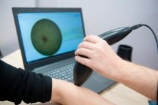
The siascopy procedure is as follows. The doctor applies a special device called a siascanner to a suspicious skin element. Within a few seconds, a multi-magnified picture of the internal structure of the neoplasm up to two millimeters deep is visualized on the monitor. The doctor has the opportunity to evaluate all detected changes without any particular problems.
The SiaScanner takes five simultaneous images, called "Siascans." The first image shows an enlarged image of the element, the second shows the vascular network, the third shows the distribution of the surface pigment, the fourth shows the distribution and concentration of the deep pigment, and the fifth shows the collagen content and the presence of additional inclusions. The doctor then collects all the information received, including anamnestic data and description, and enters it into the computer. After decoding and generating, a special program evaluates the danger of the detected skin element using a 12-point scale. [ 5 ]
After this, the doctor evaluates the results. As a rule, if the pathological suspicion index is less than six points, then we can say that the neoplasm is not dangerous: such an element is not removed, but continues to be observed in dynamics. If the indicators are high, then the patient is sent to oncologists for subsequent extended diagnostics.
The information obtained during siascopy is stored in the clinic's file. The doctor additionally makes a duplicate and gives it to the patient. If necessary, sends an electronic version to the attending specialist. The patient can go home after the procedure: no additional actions are required on his part. [ 6 ]
Contraindications to the procedure
Experts point out that there are no contraindications to diagnostic siascopy. The procedure involves the use of a regular light beam, which does not have any, even minimal, negative impact on either the skin or the entire human body. Siascopy can be prescribed to any patient, at any age, in any health condition. [ 7 ]
Consequences after the procedure
If during the siascopy examination the doctor discovers a malignant pathology, he will definitely refer the patient to an oncologist. The next step may be a histological examination, which is necessary in any case for the removal of pathological elements on the skin (especially those associated with changes in pigmentation).
If a person is diagnosed with a benign tumor and refuses to have it removed, they are given the diagnostic results and sent home. If the patient so desires, the tumor is removed. This can be done using the following methods:
- laser removal;
- radio wave removal.
The proposed methods are quite effective and leave virtually no marks or scars on the skin.
No negative consequences caused by the siascopy procedure itself have been detected. [ 8 ]
Complications after the procedure
Siascope is one of the most accurate, effective and safe diagnostic methods in dermatology. The procedure is based on spectrophotometric intracutaneous analysis. A special device called Siascope, developed by employees of the University of Cambridge, is used for diagnostics.
Siascopy is absolutely safe and does not cause complications or negative consequences. The essence of the procedure is the interaction of light waves of different lengths with the pigment substance melanin, as well as with collagen and hemoglobin. [ 9 ]
There are no restrictions on the number and frequency of siascopy studies. Diagnostics can be performed at any age, as well as during pregnancy and breastfeeding.
Care after the procedure
Since siascopy is a non-invasive and safe procedure, the patient does not require additional time and resources for recovery. He can return to work and lead a normal life immediately after the examination. However, it is necessary to strictly follow the doctor's recommendations. For example, if a malignant tumor is detected during siascopy, the patient is urgently referred to an oncologist, where he undergoes a number of additional examinations and is prepared for tumor removal. [ 10 ]
If a benign tumor is detected, the doctor may recommend removing it. However, if the patient does not feel any discomfort associated with the presence of the tumor, then there is no need to rush to remove it, but to observe it dynamically. At the same time, the patient will be given the following recommendations:
- protect the skin from ultraviolet radiation, avoid intense sun exposure;
- do not visit a solarium;
- avoid damage and injury to the skin in the area where the pathological element appears;
- regularly examine the skin, monitor the growth of the neoplasm, periodically visit a dermatologist to monitor the growth of the pathology;
- If signs of malignancy appear (discharge, bleeding, scales and crusts, pain, tingling, swelling), you must consult a doctor.
Reviews
Thanks to siascopy, dermatologists have managed to significantly reduce the number of diagnostic errors and increase the rate of timely removal of skin lesions. In general, doctors note the following positive aspects of siascopy:
- allows you to detect skin pathology in literally half a minute;
- can save diagnostic results and, if necessary, compare indicators;
- facilitates dynamic monitoring of suspicious elements;
- allows for a comprehensive analysis and assessment of the state of the neoplasm;
- Siascope is lightweight, portable and convenient, and the procedure is comfortable for both the patient and the doctor;
- the method is diagnostically accurate.
It is very convenient that the scans after siascopy can be saved in memory and, if necessary, used later by superimposing or visual comparison during repeated consultations. A special programmer creates a reference point, and the indicators are exported to the entered e-mail address. In this way, you can even get a conclusion remotely. [ 11 ]
It is important that siascopy is able to examine suspicious elements at the initial stage of their development, when they are still invisible to the naked eye.