Medical expert of the article

New publications
Squamous epithelium in smears in women and men
Last reviewed: 04.07.2025

All iLive content is medically reviewed or fact checked to ensure as much factual accuracy as possible.
We have strict sourcing guidelines and only link to reputable media sites, academic research institutions and, whenever possible, medically peer reviewed studies. Note that the numbers in parentheses ([1], [2], etc.) are clickable links to these studies.
If you feel that any of our content is inaccurate, out-of-date, or otherwise questionable, please select it and press Ctrl + Enter.
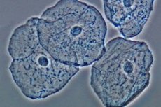
Flat epithelium in a smear is a medical concept used by doctors examining the health of the reproductive system of women and men. Epithelial cells in biomaterial are detected in a laboratory, and their number can tell a specialist both about the normal state of the genital area and about various pathological processes occurring inside and outside the human body. To obtain such information, a person must take a smear for cytology, which is often called a smear for microflora.
What is squamous epithelium?
For a gynecologist or urologist, flat epithelium in a smear for microflora is an important source of information, despite the fact that the doctor himself cannot examine these microparticles when taking a smear. Cells and layers of epithelium are detected during the study of biomaterial taken from the surface of the cervical canal and from the walls of the vagina in women or from the urethra in men, under a microscope.
Almost our entire body is covered with a protective membrane that we call skin. But the skin in the oral cavity, around the eyeball, in the vagina, in the urethral canal, etc. is somewhat different, since it is not made of such a strong material. Such delicate skin, covered with mucous secretions, is usually called the mucous membrane, and its superficial layer, which covers the epidermis, is called the epithelium.
Despite the fact that the mucous membrane is abundantly supplied with blood vessels, which explains its bright pink or red color, there are no such vessels in the epithelium. The nutrition of epithelial cells is carried out through the basement membrane.
Despite the fact that the thickness of the epithelium does not exceed 150-200 microns, this covering of internal organs is considered multilayered, i.e. the cells in it are arranged in several layers. Flat epithelium is precisely the closest superficial layer of the mucous membrane, consisting of flat epithelial cells.
There are 3 types of epithelial cells: superficial, intermediate and basal, located at different levels. The lower layer closer to the epidermis is called basal, and a layer of cylindrical (basal) cells is attached to it, performing a protective function.
But our body is constantly moving and undergoing renewal, which also concerns epithelial cells. Basal cells in the process of division (proliferation) form a layer of cells (intermediate cells) that have a complex shape with growths and spines and are located one above the other. Over time, these cells become flat and pass into the surface layer, which is renewed regularly once every 5-7 days. Old cells at the very surface of the epithelium are exfoliated and come out together with mucus and other physiological secretions.
It is this dead flat epithelium that doctors subsequently discover in a smear, separated from the main mass. It would seem that there is nothing surprising or pathological about this, because it is a natural process of cleansing and renewing the mucous membrane. In fact, everything depends on the number of epithelial cells detected, and both an increase and a decrease in their number compared to the norm are considered dangerous.
Flat epithelial cells are found in smears in both men and women, regardless of whether their genitourinary system is in order. Since epithelial renewal is considered a physiologically determined process, it is not surprising that some amount of flat epithelium will be found even in a healthy organism.

Tests: How to Prepare Properly
Sometimes flat epithelium in a smear may indicate not so much a disease or some non-pathological changes in the body, but rather improper preparation for the test or improper smear collection. A gynecologist or urologist may prescribe a microflora test if the patient has contacted him about pain in the pelvic area, redness and swelling of the external genitalia, unusual discharge, as well as symptoms such as burning, itching, pain during urination or sexual intercourse.
Such studies may be prescribed as part of a routine medical examination or when planning a pregnancy. In parallel, a urine test is usually prescribed, which helps to identify hidden diseases of the entire urinary system. But they can also manifest themselves in the form of the appearance of epithelial cells and leukocytes in the urine, while a smear only allows us to judge the inflammation at the site of its collection (urethra, vagina or cervical canal).
But no matter what analysis is prescribed, before it is carried out, it is necessary to carry out hygienic procedures, i.e. thoroughly wash the external genitals with water without using soap or other hygiene products. If a smear is planned to be taken from the vagina, women think that it is necessary to thoroughly wash everything inside using douching. In fact, this should not be done, since the results of the analysis will be distorted (it will not show the real content of leukocytes and epithelial cells separated from the walls).
To ensure reliable smear results, it is recommended to abstain from sexual intercourse and use of contraception two days before the procedure. A visit to the toilet for a small need should occur no later than 1.5-2 hours before the procedure.
Gynecological examination and smear testing are of no value if they are performed during menstruation. However, it is believed that the examination will be more reliable if it is performed during the first week after the end of menstruation. In the second phase of the menstrual cycle, the results will be completely different, and pathology can simply be overlooked.
Proper preparation for the test will help the doctor make a reliable diagnosis, and the patient will not be upset in vain if the test shows an inaccurate result due to non-compliance with the above requirements, which is what happens most often. The reason for a large number of unreliable results is the lack of knowledge about preparation for tests and the haste of doctors, who often take a smear without preliminary preparation.
 [
[ Who to contact?
Non-pathological causes of changes in the amount of squamous epithelium in a smear
The norm of flat epithelium in a smear differs slightly depending on the patient's gender. A smear from the vagina or cervical canal in women should contain visible epithelium in the amount of 5-15 units. The difference in numbers is explained by the fact that the renewal of the surface layer of the epithelium does not occur daily, therefore, at the time of cell renewal, more of them are detected in the smear, and during breaks, the number of dead cells decreases (they are excreted from the body naturally).
A urogenital smear in men should show 5 to 10 units of epithelial cells. Since the smear is taken from the urethra, which also serves as the urinary canal, it should be taken into account that some of the epithelial cells will be washed out by urine, so epithelial cells in the amount of 13-15 units can already indicate pathological processes in the patient's reproductive system.
As we have already mentioned, the renewal of epithelial cells is considered a normal natural process, so dying cells should be present in the urogenital smear. Their absence or insufficient quantity is as alarming a factor as an increase in the number of mature epithelial cells with a small nucleus and large cytoplasm separated from the surface of the mucous membrane. But it is important to understand that a change in the number of epithelial cells in a smear does not always indicate a serious pathology.
Risk factors for an increase or decrease in the amount of squamous epithelium in the vagina or urethra may include:
- Drug therapy and contraception. The quantitative indicators of squamous epithelium in the smear may be affected by the use of oral contraceptives and hormonal agents, including anti-inflammatory drugs from the glucocorticosteroid category.
- Climax in women. A decrease in estrogen production and age-related changes in the female reproductive system lead to the fact that the amount of squamous epithelium in the smear gradually decreases. In the pre-climacteric period, epithelial cells in the material taken from the surface of the vaginal mucosa or cervical canal can be found in the amount of 1-3 pieces (a single squamous epithelium in the smear), and at the onset of menopause, they can be completely absent.
- Menstrual cycle phases. The hormone estrogen is responsible for the renewal of cells in the internal genital organs of women. When its production is at its maximum (the middle of the menstrual cycle), the number of squamous epithelial cells in the smear increases. In the second half of the menstrual cycle, the body prepares for possible conception, so the production of the hormone progesterone, responsible for the normal course of pregnancy, increases. Against this background, the processes of renewal of the vaginal mucosa subside a little and the number of epithelial cells in the smear decreases slightly.
- Piercing in the intimate area.
As we can see, the number of squamous epithelial cells in a woman's body can be used to judge her hormonal background. Any disturbances will be reflected in the test results even before the first signs of a disease caused by hormonal imbalance appear.
Increase or decrease of squamous epithelium in a smear as a symptom of disease
So, a smear cytology analysis can show normal (in the range of 5-15 units), increased or decreased content of epithelial cells in the material taken for examination from the urethral canal or internal genital organs. If the number of flat cells is within the normal range, there is nothing to worry about. A correctly taken smear should contain flat, cylindrical and glandular epithelium, which lines the mucous membrane of the internal organs, but within the normal range.
But indicators above or below the normal limits should already alert the doctor and the patient, because they can indicate some pathological processes occurring in the body of a man or woman. And the sooner these processes are identified, the less harm they will cause.
Squamous epithelium in a smear in women
Most often, epithelium in large quantities indicates an inflammatory process localized in the area of the internal genital organs:
- Vaginitis (inflammation of the vaginal tissues),
This fairly common disease usually does not proceed asymptomatically, so squamous epithelium in a smear in large quantities is not the only manifestation of the disease. Usually, a smear with such indicators is taken when a woman consults a doctor with complaints of:
- discomfort in the area of the internal genital organs, pain during sexual intercourse,
- itching in the genital and vaginal area,
- irritation of the external genitalia, resulting in redness and swelling of the labia, a burning sensation that intensifies during urination,
- the appearance of copious discharge, which is white or yellowish in color, and there is so much discharge that the woman feels constant wetness in her panties, which causes irritation of the external genitalia.
The main cause of vaginitis is pathogenic and opportunistic microflora that has entered the vagina and begun to actively multiply due to an imbalance in the microflora of the internal genital organs, mechanical damage, hormonal disorders, etc. As for the discharge, its nature (color, odor, and other indicators) will depend on the type of pathogenic microorganisms inhabiting the vagina (gonococcal or trichomonas infections, activated opportunistic microflora, fungi, etc.). If the cause of the disease is gardnerella, in addition to squamous epithelium, key cells will be detected in the smear. Since vaginitis is an inflammatory disease, the number of leukocytes will also be increased.
- Cervicitis (inflammation localized in the cervical canal of the cervix),
This inflammatory disease can be overt or latent, so the doctor may accidentally detect increased superficial squamous epithelium in a smear during a routine medical examination. If the pathology is acute, the woman will indicate the following symptoms:
- quite abundant mucous or purulent discharge from the vagina (their nature again depends on the pathogen, which can be bacteria or viruses, as well as fungi, for example, actinomycetes),
- dull pain in the lower abdomen that is not constant.
Examination on a gynecological chair will show redness and swelling of the tissues near the entrance to the cervical canal. If the cause of the disease is gonococci, the inflammatory process and its symptoms will be especially pronounced, while chlamydia provokes a sluggish inflammation with less noticeable symptoms.
Trichomonas severely damage the mucous membrane, causing microscopic hemorrhages to appear on it. They also contribute to the appearance of modified cells in smears. Therefore, when flat epithelium without atypia is detected in a smear, trichomonas infection can be excluded. But it should be remembered that again, it is not so much the quality of epithelial cells that is important, but their quantity, although the presence of atypical cells can help the diagnostician suspect trichomonas in the development of the disease, which should subsequently be confirmed by bacterial analysis.
But if the unchanged flat epithelium in the smear is alarming, if it appears in quantities exceeding the norm, then what can we say about cells with an atypical structure. The presence of atypical cells does not necessarily indicate a trichomonas infection, but this is not a reason to relax. After all, modified epithelial cells can also indicate the development of an oncological process, so a more detailed study is required. If atypical cylindrical epithelial cells are detected, then we may be talking about cervical cancer.
In the case of viral cervicitis, the herpes virus or human papillomavirus is usually the first suspected cause. The herpes virus makes the surface of the cervix bright red and loose in appearance, transparent bubbles may appear on it, which, when bursting, leave behind small red wounds (ulcers). In the case of papillomatosis, ulcers may also be seen on the tissues of the cervix, and sometimes pathological growths ( condylomas ) form on it.
If the cause of inflammation of the tissues of the cervix is radiant fungi ( actinomycetes ), a yellowish granular coating is found at the site of the lesion, and Candida fungi leave behind a white mass resembling cottage cheese (grainy and with a sour smell), which is very easily removed from the reddened surface.
A similar coating is observed in leukoplakia (hyperkeratosis), a disease in which keratinization of the mucous membrane occurs (normally, keratinization processes of the mucous membrane are not characteristic). But in this case, unlike a fungal infection, the whitish or grayish coating (it can form on the cervix and on the vaginal mucosa) is difficult to remove, and the smear reveals squamous epithelium scales, which are the cytoplasm of a cell without a nucleus.
By the way, sometimes doctors find not individual cells in a smear, but layers of squamous epithelium. If these are isolated formations, there is no need to worry too much, because the cells of the epithelium of the uterus and vagina are arranged in layers, so it is not surprising if they separate from the total mass in a whole section during cell renewal. But if there are many such layers, this may be evidence of dysplastic processes in the uterus (dysplasia, erosion, endometriosis, leukoplakia of the cervix, precancerous conditions and cervical cancer), so further examinations and consultation with a gynecologist, and possibly an oncologist, are required.
Acute cervicitis is characterized by the presence of a large number of leukocytes in the smear (leukocytosis), lymphocytes and histiocytes appear, and cylindrical and squamous epithelium are detected. In this case, the cells of the cylindrical epithelium (basal layer) have an enlarged nucleus, and the squamous epithelium of the superficial layer in the smear has dystrophic changes (for example, scales appear that do not contain nuclei).
If the disease has been progressing in a sluggish form for a long time, smears will show a large number of columnar epithelial cells of different sizes and signs of destruction of cellular structures.
- urethritis (an inflammatory process in the tissues of the urethra, which can be diagnosed in patients of both sexes).
Urethritis is a disease that can have different origins. The infectious form of the disease is an inflammation of the urethral walls caused by bacteria. Non-specific urethritis is said to be caused by the proliferation of opportunistic microflora. This includes staphylococci, streptococci, E. coli and other representatives of this group.
Specific urethritis is a disease caused by sexually transmitted bacteria ( gonococci, trichomonas, chlamydia, mycoplasma, gardnerella, etc.) or fungi of the genus Candida. Sometimes the cause of inflammation of the internal tissues of the urethra are herpes viruses and human papillomaviruses (viral urethritis).
Non-infectious urethritis is usually a complication of existing diseases: cystitis, urolithiasis, tumors along the urethra, injuries (often this happens when inserting a catheter, during cystoscopy, etc.), congestion in the blood vessels of the pelvic area, allergic or gynecological diseases. Non-infectious urethritis can even be provoked by the first sexual experience.
Chronic urethritis in 50% of cases occurs without any symptoms that bother the woman, and it can only be detected using a smear taken from the urethra. Most often, urethritis in women occurs against the background of cystitis, which is why they consult a doctor. Often, cystitis is a complication of urethritis, which occurs in a protracted form, especially if we are talking about the infectious nature of the pathology.
Acute urethritis is characterized by:
- discomfort when urinating (a woman may experience itching, burning, a feeling of squeezing or tightening of the tissues of the urethral canal),
- irregular aching pain in the very lower abdomen,
- discharge from the urethra (not vaginal!), which, depending on the pathogen, can have a different character (mucous or purulent, which is why in the morning there is an unpleasant sensation as if the tissues of the urethra are stuck together),
- the appearance of blood particles in the urine as a result of severe irritation and ulceration of inflamed tissues,
Another feature of urethritis is that the patient does not experience symptoms of general malaise. There are mainly local symptoms. And the disease can be detected by chance, by detecting flat epithelium in the urine, where it should not be, or an insignificant number of separated epithelial cells that previously lined the organs of the urinary system (no more than 3-4 units) can be observed.
Flat epithelium in urine is quite often found during pregnancy, when the woman's body, and especially the urinary system, experiences excessive stress. The appearance of a large number of separated epithelial cells indicates, first of all, an inflammatory process occurring in the present or past tense.
If we exclude gynecological problems (and the proximity of the urethra and vagina means that the secretion of both organs can mix, so it is very important to properly prepare for urine analysis and smears), the cause of an increased number of epithelial cells in the urine can be:
- cystitis (inflammation of the walls of the bladder, which also has a surface layer of epithelial cells inside, which as a result of inflammatory processes can peel off from the walls of the organ),
- urethritis (inflammation of the urethral mucosa, which we have already written about),
- nephropathy of pregnancy - a manifestation of late toxicosis with increased blood pressure, edema syndrome and the appearance of protein in the urine, epithelial cells of various layers of the kidneys, erythrocytes, etc.
Both during pregnancy and at other times, the appearance of components in the urine that are not typical for it can provoke nephropathy caused by improper functioning of the immune system, taking medications, some endocrine diseases (for example, diabetes mellitus ), etc., in which a large amount of squamous epithelium is found in the urine.
It should be said that due to inflammatory processes in the kidneys and bladder, flat epithelium in urine can be found not only in women but also in men. It is clear that in smears from the urethra, the doctor can again detect such cells.
Squamous epithelium in a smear in men
In men who usually consult a urologist about various discharges from the penis, as well as such unpleasant symptoms as itching, burning and inflammation of tissues in the area of the urethra, a smear for microflora is taken from the urethra. In addition to bacteria, viruses, fungi and other microorganisms, exfoliated cells of flat and cylindrical epithelium can also be found in the mucous, whitish or purulent discharge. The mucous membrane of the urethra has a slightly different structure, so a small number of basal cells is not a pathology.
If the number of epithelial cells in the smear is higher than normal, we are talking about tissue destruction. A large number of squamous epithelium, as in the case of women, most often indicates an inflammatory process in the genitourinary system. In this case, it is necessary to take into account not only the urinary organs (kidneys, bladder, urethra).
The inflammatory process can also be localized in the seminal sacs (testicular inflammation or orchitis ) or the prostate gland (prostate inflammation or prostatitis ), which are classified as genital organs. And since the organs of the urinary and genital systems have a common outlet (the urethra), there may be more reasons for discharge from the penis in men than in women, whose outlets, although located nearby, are still separated, which facilitates diagnosis with the correct smear.
If we are talking about an inflammatory process, smears will also necessarily show an increase in the level of leukocytes (more than 5 units in the field of view), which are released by the immune system to fight the infection. Bacteriological studies also reveal pathogens, which once again suggests that the appearance of squamous epithelium in the smear is not accidental.
If neither infection nor leukocytes are found in the smears, then we are talking about hyperkeratotic processes (leukoplakia, etc.), which can be localized in various areas of the mucous membrane throughout the body. But it is important to understand that the amount of squamous epithelium can increase slightly compared to the norm as the human body ages. Although this process is not so clearly expressed in men, doctors still note some changes in smear tests that are not associated with diseases of the genitourinary system.
In both women and men, the increase in the number of epithelial cells in the smear may be temporarily increased due to the use of local agents. In this case, the situation can be considered an unusual manifestation of an allergic reaction to the drug. By analogy, itching, redness and peeling of the skin may appear, which is also explained by the rejection of superficial epithelial cells.
As for the reduced number of epithelial cells in the smear, this is considered an alarming symptom for young women. With the onset of menopause, a decrease in the production of the female hormone estrogen leads to a decrease in such cells in the tests, which is considered a physiologically conditioned age-related process. Hormonal imbalance towards a decrease in estrogen production in young women before menopause is fraught with big troubles in the form of a violation of the internal environment of the vagina and the activation of pathogenic microflora.
With estrogen deficiency, gradual atrophy of vaginal cells occurs, which manifests itself in the absence of natural ovulatory discharge, vaginal dryness during intercourse, slight vaginal spotting, burning and itching in the vagina, frequent urge to urinate. All these are quite dangerous symptoms that can have tragic consequences and cause infertility in women of childbearing age. Therefore, it is undesirable to ignore such symptoms.
Consequences and complications
In a smear for microflora, you can find different types of epithelium: flat superficial, transitional, cylindrical, which lines the cervical canal and is periodically displaced by flat epithelial cells. There is nothing pathological in the fact that these cells are present in the biological material. This is the result of physiological processes occurring in the body. Women may have fewer of these cells, women may have more, but this is also due only to the structure of their genitals. And if the number of cells of a particular type when studying biomaterial under a microscope does not exceed 15 units in the field of view, there is nothing to worry about.
But if the indicators are too high or below the norm, this is already a reason to specifically take care of your health. Especially when it comes to women of reproductive age.
An increased amount of squamous epithelium in a smear most often indicates inflammatory processes in the urethra or vagina - organs whose surface is covered with a delicate mucous membrane. And as we know, no inflammatory process, especially a long-term one, passes without a trace. Timely treatment of an acute inflammatory process, which can be indicated by a large number of cells of exfoliated squamous epithelium, helps prevent possible complications.
For example, the transition to a chronic form of vaginitis threatens in the future with inflammatory processes of the external genitalia, erosion of the cervix, inflammation of the endometrium, which in turn can eventually lead to dysplastic processes ( dysplasia of uterine tissue, endometriosis, cervical cancer ). In childhood, vaginitis is fraught with the fusion of the labia minora and labia majora in girls.
The inflammatory process can lead to the compaction of the mucous tissue. Metabolic processes and cellular respiration are disrupted, and the mucous tissues atrophy. Cracks and erosions can appear on the mucous membrane, which not only become an obstacle to receiving pleasure during sexual intercourse, but also become a breeding ground for the reproduction of infection. If vaginitis itself is not always caused by an infection, then the appearance of various lesions on the mucous membrane will provoke its addition.
An infection, especially of a bacterial nature, has the ability to cover ever larger areas, i.e. it will move upward, affecting the organs of the urinary system.
Chronic cervicitis can have identical consequences. The infection will gradually spread to other organs, causing inflammation of the appendages, bladder, and peritoneum. It can result in the appearance of adhesions on the internal organs and the development of cancer (cervical cancer does not develop so often against the background of inflammation, but such a danger still exists). In addition, the appearance of a large number of cylindrical and flat epithelial cells in a smear can indicate the initial stage of cancer without symptoms of cervicitis.
Urethritis in women, in the vast majority of cases caused by an infectious factor, although damage to the urethra can also be provoked by urolithiasis, can cause a violation of the vaginal microflora, inflammation of the bladder and even the kidneys if the infection rises to them.
In men, untimely or poor-quality treatment of urethritis (and even more so any of its therapeutic measures) causes many unpleasant consequences that will also affect sexual life:
- development of prostatitis, and as a consequence prostate adenoma,
- the appearance of vesiculitis, orchitis and colliculitis (inflammatory processes in the seminal vesicles, testicles or seminal tubercle),
- development of balanoposthitis (inflammation of the penis in the area of the foreskin), etc.
Long-term inflammation in the urethra can lead to the appearance of strictures, i.e. to a narrowing of the urethra, which creates problems with the removal of urine from the body and often requires surgical intervention.
Patients with inflammatory diseases of the genitourinary system begin to have problems of a sexual and psychological nature. Pain during sexual intercourse becomes the reason for refusal of sexual intimacy, constant dissatisfaction of sexual partners. Quarrels and scandals begin in the family, which can also be considered a remote consequence of a careless attitude to the appearance of a large number of epithelial cells in the smear.
Although no lesser sexual problems can be observed in women with insufficient epithelium in smears, in whom the lack of lubrication in the vagina leads to pain and irritation of the mucous membranes during sexual intercourse.
But pathologies that change the composition of the smear have much more unpleasant consequences, considering that they sometimes become an obstacle to conceiving a child. And infertility of one of the spouses often leads to the breakdown of the family.
As we can see, the detection of squamous epithelial cells in a smear in quantities different from the norm can be considered not only an indicator of pathological processes in the body, but also an opportunity to promptly begin effective treatment and prevent the development of dangerous complications.
Prevention and prognosis
When it comes to human diseases, the prognosis for their treatment depends on many factors, so it is never 100%. Flat epithelium appears in a smear taken from the genitals of men and women, depending on its quantity, it can indicate various pathologies caused by hormonal disorders, inflammation, infection, failures in the immune system, etc. It is clear that treatment in each specific case will be considered individually, and its effectiveness will depend on the competence of the doctor.
But the prognosis of treatment is influenced not only by the prescribed treatment. The time of seeking help also plays a major role. The earlier the disease is detected, the greater the chances of saying goodbye to it without consequences and complications. This applies to both inflammatory pathologies and oncological diseases, where it is not even about health, but about the patient's life.
It is clear that compliance with the doctor's requirements will certainly affect the treatment prognosis. If the patient did not want to undergo a full course of treatment, one usually cannot count on a good, lasting result. There is a high risk that the disease will return after a while.
Let's not go into too much detail about the consequences that a person may face if he does not take any measures to treat the disease. Let's talk about how to make the treatment more effective.
When treating infectious and inflammatory diseases of the genitourinary system, it is very important:
- Maintain hygiene of the genitals. You will have to wash even more often than usual, especially during menstruation.
- You will also have to change your underwear and intimate hygiene products (tampons, pads) more often.
- All medical procedures should be carried out after hands have been thoroughly washed with soap. Hands should also be washed after medical procedures.
- The genitals, where the infection is hidden, require care using a separate towel. It is better if these are disposable napkins. Otherwise, the towel will have to be washed and ironed with a hot iron every day.
- Particular attention should be paid to underwear. It should be made of natural fabrics, "breathable", because the "greenhouse" effect created by synthetics only contributes to the proliferation of pathogenic microflora.
- During treatment of the disease, sexual contact should be avoided. This will help prevent secondary infection and the spread of the infection to other people, in particular to the sexual partner. Failure to comply with this requirement is one of the reasons for the prevalence of sexually transmitted diseases. Some patients believe that if they have started treatment, they are no longer contagious. Unprotected sex is especially dangerous.
- It is important to understand that if two people regularly have sex and one of them is diagnosed with an infectious disease, both partners should be treated. Even if we are not talking about sexually transmitted diseases, such as candidiasis or vaginitis caused by opportunistic microflora, some preventive measures in the form of treating the penis with antiseptics that are effective against bacteria and fungi will not hurt. Especially if you consider that not everyone can boast of a strong immune system.
- The treatment procedures prescribed by the doctor must be carried out regularly and in full. You cannot stop treatment just because the symptoms of the disease disappear. You need to complete the full therapeutic course and then, if necessary, resort to preventive measures.
But any disease is easier to prevent than to cure. This is the truth that you need to be guided by in your life, so that there are fewer unpleasant moments in it.
To avoid infectious and inflammatory diseases of urogynecological nature, you don't need much. You should pay attention to your sex life. Even if your sexual partner is regular, you should carefully monitor your health. After sexual intercourse, carrying out hygiene procedures will help prevent possible infection.
If we are talking about casual sexual relations, it is necessary to use high-quality personal protective equipment (condom). And again, do not forget about genital hygiene, trying to take a shower as soon as possible.
Wearing underwear made of natural fabrics will help prevent the proliferation of opportunistic microflora, which loves warm and humid places. Increased humidity, as well as allergens contained in scented pads, synthetic underwear, and some intimate hygiene products, which are best avoided, can cause irritation of delicate tissues.
As for the prevention of diseases in which the amount of flat epithelium in a smear differs from the established norm, here the most important thing is to maintain intimate hygiene, proper nutrition that provides the body with nutrients necessary for strong immunity, and caution in sexual intercourse. Maintaining the general health of the body and timely treatment of infectious diseases, regardless of their location, also play an important role. It is always necessary to remember the ability of infection to spread throughout the body with the blood and lymph flow in order to prevent such a danger in time.