Medical expert of the article

New publications
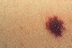
Melaniform nevus
Last reviewed: 12.07.2025

All iLive content is medically reviewed or fact checked to ensure as much factual accuracy as possible.
We have strict sourcing guidelines and only link to reputable media sites, academic research institutions and, whenever possible, medically peer reviewed studies. Note that the numbers in parentheses ([1], [2], etc.) are clickable links to these studies.
If you feel that any of our content is inaccurate, out-of-date, or otherwise questionable, please select it and press Ctrl + Enter.
A nevus or birthmark is a skin formation made up of modified cells of the skin pigment melanin. It is defined by Russian dermatologists as a melaniform nevus, which can be acquired or congenital. [ 1 ]
On the issue of terminology and classification
The part of the definition “form” (from Latin – formis) in clinical terminology means “similar, similar”.
In the Russian version of ICD-10 (in the section on benign neoplasms), melaniform nevus (pigmented, hairy and blue) has the code D22. Also, depending on the localization, melaniform nevus of the lip has the code D22.0; nevus of the eyelid is coded D22.1, and of the ear - D22.2; melaniform nevus of the scalp and neck - D22.4; melaniform nevus of the face (its unspecified parts) - D22.3; melaniform nevus of the trunk - D22.5.
In addition, a mole located on the peripheral parts of the body can be defined as an acral melaniform nevus, and a nevus on the upper limb has the code D22.6, and on the lower limb - D22.7.
There is also a term "melanocytic nevus". And the question arises, what does melaniform and melanocytic nevus mean, what is the difference between them?
The fact is that in the original English-language classification – International Classification of Diseases (ICD 10) – nevus is defined as melanocytic (the term “melaniform” does not exist at all). And melanocytic nevus is the same mole on the skin, a benign neoplasm or epidermal nevus, consisting of the same melanocytes (lat. – melanocyte), which are often called nevocytes, that is, nevus cells.
Epidemiology
On average, an adult Caucasian may have between one and four dozen melaniform (melanocytic) nevi, with most of the formations located on the body above the lumbar region.
During childhood and adolescence, the number of moles gradually increases, with the peak of their appearance occurring between the ages of 18 and 25. [ 2 ]
Causes melaniform nevus
The main causes and possible risk factors for the appearance of melanoma nevi are discussed in detail in the publications:
Melaniform nevus in children of the first year of life is rarely observed (on average, in 5-7% of infants). [ 3 ] Read:
Pathogenesis
The process of nevi formation – nevogenesis – is explained by a complex interaction of genetic factors and environmental factors (exposure to ultraviolet radiation, etc.).
The pathogenesis of congenital nevi is associated with disturbances in the formation of melanocytes from precursor cells – melanoblasts of the neural crest of the embryo. Dendritic melanocytes, which transfer pigment to the surrounding keratinocytes of the skin, and nevus cells (nevocytes) are morphologically different types of cells. [ 4 ]
According to modern models of nevogenesis, melanocytic skin lesions arise from a single mutated cell, the activation of which leads to the proliferation of melanocytes and their transformation into nevocytes. Pigmented nevus cells in the upper dermis and epidermis have an epithelioid appearance with a cuboid or oval shape, diffuse cytoplasm, and a round or oval nucleus; nevocytes in the middle dermis are smaller in size and do not contain melanin, and nevus cells in the lower dermis are spindle-shaped, like fibroblasts.
In this case, the formation of nevi is associated with mutations in the genes of the signaling protein NRAS (which is involved in the regulation of mitosis – cell division); proteins BRAF (serine-threonine kinase), FGFR-3 (fibroblast growth factor receptor), etc.
And these changes at the gene level affect transcription factors and signaling pathways (cellular transduction) in such a way that the proliferation and differentiation of melanocytes is disrupted. [ 5 ]
Symptoms melaniform nevus
A growth on the skin is the first sign of a melaniform (melanocytic) epidermal nevus. It can be nodular, located at the junction of the epidermis and the underlying dermis - in the dermoepidermal layer; it can be slightly raised above the surface of the skin, as well as intradermal, which is a classic birthmark - a raised dome-shaped bulge on the surface of the skin from light brown to dark brown in color, of varying degrees of convexity and size, round or irregular in shape.
There are different types of melaniform nevus, that is, types of moles on the body, the external symptoms of which may differ.
Melanocytic or melaniform pigmented nevus is an acquired melanocytic nevus that appears in childhood as brown, flat spots measuring 1–2 mm in size that most often appear on skin exposed to ultraviolet rays. In these nevi, nests of nevocytes are located along the dermoepidermal junction.
If the area of the pigmented formation exceeds 10-12 mm, an extensive melanoform nevus is defined. For example, the size of Becker's nevus (hairy epidermal nevus) can reach 15-20 cm.
Intradermal melanocytic nevus or convex mole refers to acquired lesions that are localized mainly on the head, neck or upper body. In this case, the nevi may be limited to the middle layer of the dermis. These intradermal nevi are usually fleshy, may have a stalk and develop in three stages: first, the mole grows rapidly (and may form a stalk), then its growth slows down and stops, after which the formation - as the depth of the dermis increases - begins to decrease and becomes lighter.
Borderline intradermal nevus is flat, can have a color from gray to brown. Also read - Flat moles
The nevus of Setton is characterized by the appearance of a depigmented ring around the mole, while the nevus of Jadassohn-Tiche (blue epidermal nevus) looks like a dense papule or nodule of blue-gray or blue-black color.
For more information on what a melanomatous papillomatous nevus or warty nevus is, see the following materials:
A mixed melaniform nevus is determined by histology of the mole; in such cases, a combination of various nevus cells, as well as connective tissue and dermal elements, is detected in one formation.
Biopsy and histological examination may also reveal an atypical melaniform nevus – a pigmented melanocytic formation on the skin with distinct clinical and histological features in the form of cellular atypia. More details in the article – Dysplastic nevi
Complications and consequences
Complications and consequences of melanoma nevi are associated with their damage (trauma), which can lead to bleeding and/or the development of inflammation.
In addition, some moles have an increased risk of malignant transformation into melanoma, so any changes in their size, shape or color should be addressed by a doctor. [ 6 ]
For more details see:
Diagnostics melaniform nevus
Read more in the articles:
Treatment melaniform nevus
Surgical treatment, that is, surgical removal of a melanoma nevus [ 9 ] is discussed in detail in the articles:
Prevention
To date, there are no preventive measures to prevent the appearance of nevi, but it is recommended to avoid prolonged exposure to the sun and not visit solariums.
Forecast
For most people, the prognosis for a melanoma nevus is favorable, but the possibility of malignancy of these skin lesions, which are initially benign, should be taken into account.