Medical expert of the article

New publications
Papillomatous nevus of the skin
Last reviewed: 12.07.2025

All iLive content is medically reviewed or fact checked to ensure as much factual accuracy as possible.
We have strict sourcing guidelines and only link to reputable media sites, academic research institutions and, whenever possible, medically peer reviewed studies. Note that the numbers in parentheses ([1], [2], etc.) are clickable links to these studies.
If you feel that any of our content is inaccurate, out-of-date, or otherwise questionable, please select it and press Ctrl + Enter.
A mole is one of the most unusual natural decorations on the human body. Some people find it charming and consider it very cute. Others complain that a mole on some part of the body is not a very desirable and convenient gift from nature. Moreover, this gift requires special attention, since a harmless benign neoplasm can turn into skin cancer, especially if it is often subject to mechanical (or thermal, chemical, etc.) damage. And it is good if the birthmark is flush with the entire surrounding skin, when the probability of damage is relatively small. But if we are dealing with a neoplasm such as a papillomatous nevus, noticeably rising above the surface, then the risk of hearing a terrible diagnosis one not very fine day becomes much higher.
What is papillomatous nevus?
A mole is a natural mark on the body that appears in humans most often in infancy. This is what the name of the neoplasm is associated with, which has become established among the people. People who have many moles on their bodies are treated in a special way. It is believed that those who are awarded a large number of dark marks by nature from birth will be very happy in life.
But is this really so, is still a question, because moles (nevi) are not always a safe decoration, which sometimes promises not so much happiness as human tragedy. And the more moles on the body, the more careful a person needs to be in life. He has to carefully protect the skin in places where nevi are localized from various damages and active sun rays, regularly examine it for changes in the shape, color or size of birthmarks and visit a dermatologist not once a year, but with any unusual changes in neoplasms.
It is especially difficult for those who have convex moles on their bodies, which include papillomatous nevus. It must be said that this type of mole is a dubious decoration, most often found on the head and neck area. But its appearance on other parts of the body is also possible.
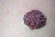
A papillomatous nevus is a cellular structure that has an irregular convex shape and is not very pleasant to look at. Such a birthmark consists of several elongated epidermal processes connected together and has a dense structure, since it is covered with a stratum corneum consisting of melanocytes. Melanocytes (pigment cells) make the neoplasm even more noticeable, although its color can vary from flesh-colored to dark brown.
When examined with a magnifying glass, a papillomatous (warty) nevus has many papillae on its surface, which is why its surface appears uneven and bumpy. Quite often, a dark brown rim can be seen around such a mole, the surface of which is flush with the rest of the skin.
As we have already said, a papillomatous nevus, resembling a strange complex wart, which is why it is sometimes called warty, is not a very pleasant-looking formation, which can cause concern in the owner of such a "decoration", who has heard about the danger of degeneration of moles into a cancerous disease called "melanoma". Therefore, many people have a question whether a papillomatous nevus is dangerous - a neoplasm with such an unusual structure and irregular shape, which is an alarming factor, because it is believed that a safe mole should have the correct shape and symmetrical structure.
Doctors believe that this type of mole, which can equally appear both at the birth of a child and throughout a person's life, is generally not dangerous. It is a benign neoplasm consisting of normal non-malignant cells, and if it is not injured, it will remain so for life. But this is where the catch lies. Any convex mole has a greater chance of being injured than its flat relatives. And if such a formation is located on the scalp, the risk of injury increases several times, because even the usual daily procedure of combing hair is an injury to the nevus.
If a mole is located on the neck, it can regularly rub against the collar, which is also considered a minor but trauma. And if a nevus receives such trauma regularly, this can lead to a change in the properties of its cells, i.e. to their degeneration into malignant ones.
As we can see, a papillomatous nevus can hardly be called an ornament due to its unsightly appearance, and its convex shape makes it not entirely safe due to the high risk of injuring the growth. It is for this reason that doctors often insist on removing such neoplasms without waiting for them to turn into melanoma.
Causes papillomatous nevus
Papillomatous nevus is one of the little-studied neoplasms on the human body. Doctors are still struggling with the reasons for its appearance, but a proven final version has not yet been put forward. Research is at the hypothesis stage, and the most logical, and therefore widespread, version is that the appearance of papillomatous nevi is one of the developmental disorders of the child in the embryonic period, i.e. in the womb.
There is an assumption that due to malfunctions in the developing organs of the embryo, a greater accumulation of melanocytes is observed in certain areas of the skin, which form a dark spot, which can have different color intensity depending on the amount of coloring pigment. Melanocytes form an intradermal nevus, which grows with the child's body and can increase under the influence of certain factors both during intrauterine development and after the birth of the baby.
As already noted, such birthmarks are observed in children from birth. It's just that in some they are more noticeable, while in others the neoplasms are practically invisible. However, during a person's life, nevi tend to increase in size several times, so at some point even a previously unnoticeable growth becomes visible.
Risk factors
Since we are talking about embryonic development disorders, there must be some reasons for such disorders, because nothing in this world happens just like that, spontaneously. Risk factors for all sorts of failures in the developing human organism can be:
- exposure of a pregnant woman to ionizing radiation (e.g. X-ray examinations, being in a radioactive zone, regular prolonged exposure to the sun, etc.),
- all kinds of poisoning (food, chemical, medicinal, toxic infections) associated with intoxication of the mother's body, because toxins are transmitted through the blood to the baby,
- infectious lesions of the genitourinary and other systems (pathogenic organisms also secrete substances that poison our body, plus stagnation in the excretory system can provoke intoxication with the products of our vital activity),
- hormonal imbalance with increased estrogen levels in the blood of the expectant mother.
Increased growth of a neoplasm in children and adults can be provoked by exposure to solar radiation (for example, a mole on the head can grow if a person does not wear hats that protect the nevus from sunlight), the use of strong chemicals at home and at work, living in an area with a high radioactive background, etc. But the growth of a papillomatous nevus does not mean its degeneration into a malignant neoplasm or other complication, if there are no suspicious symptoms (redness, inflammation, the appearance of necrotic areas, bleeding, etc.).
Pathogenesis
A papillomatous nevus is a neoplasm that resembles a wart or papilloma in appearance. Quite often, such growths are found in newborns, which is not a cause for concern. But unlike ordinary moles, the appearance of papillomatous nevi is not an alarming sign in adulthood.
In fact, warty birthmarks appear at the birth of a child, they may simply not be noticeable at first. But as it grows and rises above the skin, the neoplasm becomes more visible, especially when it is located on the face or neck. The person grows up and the birthmark grows. Usually, its growth stops after 30 years.
Papillomatous nevi, which can be intradermal or develop on the basis of an intradermal formation, are considered benign and do not belong to the group of dangerous ones with a high risk of degeneration. According to statistics, such moles very rarely develop into skin cancer, but since such a danger still exists, it should not be forgotten. Moreover, melanoma, developing at the site of localization of a once harmless mole, can have a rapid development. And in this case, it is better to remove the often injured nevus in advance and prevent the development of melanoma than to try to treat a deadly disease later.
Such growths on the skin are quite often large (1 cm and more). And large moles found in infants have a 5% tendency to degenerate into malignant neoplasms. Yes, the risk is relatively small, but it exists. And when it comes to a disease that is difficult to treat and can cause the patient's death, even a small probability becomes a big danger.
We will not frighten the reader too much, because the probability of malignancy of cells in the case of papillomatous nevus is very low. Therefore, there is no need to worry too much if the neoplasm is just a cosmetic defect with a low risk of damage in everyday life. Nevertheless, caution should become part of the patient's life, which will help prevent malignancy and other complications of warty nevus.
Symptoms papillomatous nevus
Externally, a papillomatous nevus does not really resemble a familiar birthmark. Instead of a cute dark spot, we see a bumpy bulge on the skin with a granular surface, reminiscent of a papilloma on a thick stalk (if you look at the birthmark under a microscope, you will also notice its resemblance to mulberry fruits).
But unlike a soft to the touch papilloma, a warty nevus has a denser structure and a horny layer. In addition, most often such a neoplasm is pigmented, i.e. darker compared to other areas of the skin. A flesh-colored nevus is quite rare. Most often, neoplasms have a pink, brown or even rare black shade.
Despite its irregular shape, a warty nevus has clear outlines and distinct borders. Many nevi have a dark rim.
On the surface of such an unusual birthmark, one can often find dark hairs growing directly from the neoplasm and making it look like a fibroepithelial nevus.
Nevi with or without hairs can be located not only on the scalp. They can also often be found on the neck and face. Less often, neoplasms choose the armpits, areas of skin under the mammary glands, the groin area, etc. as their location.
As for the number of such neoplasms, they can be single or multiple, scattered throughout the body or located nearby in a limited area. And if these growths did not grow, a person would perceive them as ordinary moles.
But papillomatous moles tend to grow slowly and increase to large sizes, so on the body such formations look rather unsightly, leading the owner of the moles to despondency and even depression.
A papillomatous nevus is a non-inflammatory formation, so there should be no swelling or redness around it. On the surface of the neoplasm, which has a non-uniform, bumpy shape, there should be no purulent ulcers or bleeding cracks. The appearance of such symptoms may be the first sign of inflammation and infection of the nevus due to damage from a comb, collar, nails, etc., or indicate the development of a malignant process, which is much less common.
Various changes in a mole may indicate malignancy of the cells:
- the nevus changed its color to a darker one,
- cracks and purulent-inflammatory lesions appeared on it,
- the shape of the neoplasm has changed,
- the edges of the growth became uneven and unclear,
- dots of a different color appeared on the surface of the mole, dark areas of necrotic tissue, hairs began to fall out (if they were there before),
- the skin on the site of the mole began to peel and there is itching,
- the mole became painful,
- other multiple irregularly shaped neoplasms have appeared (usually a papillomatous nevus grows until the age of 30; if similar neoplasms appear later, this is already a reason to visit a dermatologist and undergo an appropriate examination, possibly even with the involvement of more than one specialist).
It is important to understand that a rapid increase in the size of any mole, the appearance of itching, swelling, pain, bleeding and other unusual symptoms is a pathological process, because nevi are considered non-inflammatory neoplasms, and such behavior is unusual for them. The appearance of such symptoms may indicate both damage and infection of the mole, and more tragic consequences - the initial stage of melanoma.
Despite the low risk of degeneration into a malignant neoplasm, any changes in the appearance and sensations at the site of the mole should not be ignored. This will help to avoid life-threatening complications.
Forms
So far we have talked about papillomatous nevus as a certain cellular structure rising above the patient's body. But we have mentioned that such moles may differ somewhat in appearance, which makes it possible to classify them, dividing them into separate types and forms.
A papillomatous intradermal nevus, or warty neoplasm, is a formation that strongly resembles a papilloma or wart. It rises noticeably above the skin, but is almost identical in color or has a light brown or pale pink tint. It has a characteristic rough surface, and often consists of two or more lobes (several moles located close to each other in a small area). Such a mole grows very slowly, becomes inflamed and bleeds in isolated cases, and is almost never prone to degeneration into a malignant tumor.
Papillomatous and warty nevi are not different types of moles. Warty nevus is a type of warty mole that has a convex shape and a bumpy structure. Warty nevus can also be keratotic (a very dense formation with a thick horny layer) or ichthyosiform (growths similar to papilloma, sometimes have blisters on the surface, then they are called bullous).
Papillomatous melanocytic nevus is a pigmented type of convex neoplasm that can be either intradermal or borderline. Such moles most often appear on exposed skin areas (on the neck, face, under the mammary glands, etc.) that are not covered with hair. Their color is determined by the large number of melanocytes in the middle and superficial layers of the skin, so the nevus can be either brown or almost black.
Melanocytes are cells that are found in large numbers on the surface of a papillomatous pigmented nevus and are more prone to degeneration. Their presence in the upper layers of the skin makes these cells more sensitive to the negative effects of sunlight and mechanical injury, which sometimes leads to malignancy of nevus cells (albeit extremely rarely). This is more typical for border nevi with melanocytes localized in the epidermis.
The hairy type of papillomatous nevus is characterized by the fact that one or more fairly long hairs can be found on the surface of such neoplasms. Moles with hair growing on them are considered completely safe. But if a person pulls out the hairs growing on a mole, he will injure the cells of the neoplasm, and they can behave in an unnatural way in this case. The development of melanoma can also be indicated by a situation when the hair on the mole falls out on its own and does not appear again.
This type of papillomatous mixed nevus is sometimes called complex, since nevocytes (germ cells of a mole) can be located both in the middle layer of the dermis and on the border with the epidermis. If such a neoplasm consists of several nearby moles, then some of the papillae on it may have a lighter shade. The surface of such a nevus is even more uneven.
In addition to the various types of papillomatous nevus, there are also 2 forms of such neoplasms: organic and disseminated.
The organic (localized) form of warty nevi is considered more common. In this case, we are talking about both single neoplasms and several moles located close to each other on a limited area of the body. Moles can be of the same color or have different colors, even bipigmented or grayish. This form is considered safe due to the fact that it does not indicate any health problems in a person.
The disseminated (systemic) form is characterized by the appearance of multiple pigmented nevi on the body, resembling warts. Moreover, moles are located on different parts of the body. This form does not always indicate the danger of health problems, but it is still quite often evidence of serious neurological problems. Multiple moles can be observed on the body of patients with lesions of the central nervous system, and in particular with epilepsy.
Nevi in the systemic form may behave inappropriately. For example, they may grow throughout the patient's life or dry up and fall off without any outside influence.
 [ 17 ]
Complications and consequences
Although the papillomatous nevus itself does not pose a health hazard, and in most cases it is considered an unaesthetic cosmetic defect, one should not treat the neoplasm carelessly. The fact that it is rarely prone to developing into melanoma does not mean that this is impossible at all. The degeneration of a papillomatous nevus, like the malignancy of an ordinary mole, can be provoked by a common household injury (the same combing of hair), and if it is repeated regularly, it is unlikely that everything will go without consequences, especially if we are talking about a pigmented nevus.
But a certain risk of developing melanoma is not the only danger that a generally harmless "wart" may conceal. Severe damage to a nevus when combing and washing the head, during hygiene procedures, when irritated by the collar of clothing or by pure chance with the formation of a bleeding wound is fraught with the fact that an infection can get into the blood, which will cause inflammation and suppuration of the tissues of the nevus. The entry of a bacterial or viral infection into the bloodstream is also fraught with the generalization of the process (sepsis), which leads to disruption of various organs and systems of the body.
But that's not all. Unlike a cute birthmark, a papillomatous nevus cannot be considered a cute and attractive decoration on the body. Many people who are "lucky" enough to have such a gift of nature are very worried about the unattractiveness of this cosmetic defect. A brown or black wart on the face, and even with dark hairs growing out of it, is unlikely to decorate anyone.
Large pigmentless or pigmented neoplasms are visible even on the scalp if a person's hair is not thick enough. The appearance of such a mole in adolescence and adulthood can be a shock for a person. A nevus that has a strange appearance and resembles a malignant mole will cause strong emotional experiences, sometimes reaching depression.
Lack of self-confidence due to a cosmetic defect, together with worries about the safety of the neoplasm, are a strong blow to the psyche, especially in adolescence. A teenager does not yet think about the fact that a nevus can be safely removed by contacting a medical institution. Young people at this age tend to hide their problems and try to solve them on their own, for example, by removing a nevus at home in the absence of the necessary research, without observing sterility using questionable methods, which is fraught with complications not only psychological, but also physical.
Sometimes medical removal of a papillomatous nevus is the only correct solution to the problem. And not only when the mole is constantly subject to injury, but even in cases where the presence of such a cosmetic defect causes serious psycho-emotional disorders and pushes people to rash actions.
Diagnostics papillomatous nevus
No matter how carefully we describe the appearance and symptoms of a papillomatous nevus, it will not be so easy for a non-professional to determine that this is the neoplasm in front of you. The similarity of a warty birthmark with other growths and modified ordinary nevi makes diagnostics very difficult. When such a neoplasm is detected, especially if it appeared in adulthood, you should not rely too much on your own strength. It is better to spend several hours going to the doctor than to study a strange bump in front of the mirror every day and worry about its safety, inconvenience, unaesthetics, etc.
Some patients believe that as long as a mole does not bother or hurt, everything is fine with it and there is no reason to visit a dermatologist. But you should always remember that melanoma can also begin painlessly, and when pain appears, time will have already been lost. So it is better to play it safe and immediately show the strange neoplasm to a specialist. Especially since diagnostics will not take much time and effort.
Regarding various skin neoplasms, it is customary to consult a dermatologist. An experienced doctor can often tell us what we are dealing with and how dangerous the mole is, even without special studies and tests. But to make sure the initial diagnosis is correct, doctors prefer to conduct instrumental diagnostics.
Instrumental diagnostic methods that are relevant when examining moles on the body include:
- Dermatoscopy is the most popular method of studying moles using a special powerful microscope that displays an image on a computer screen (dermatoscope). It helps to see the smallest changes on the surface of the neoplasm.
- SIAscopy is a non-invasive study that involves spectrophotometric scanning of pigmented lesions using a device called a SIAscanner. This method is sometimes called deep dermatoscopy, because it allows you to get a three-dimensional image of the dermatoscopy result, melanin and collagen of the skin, blood. The accuracy of the study in detecting melanoma-hazardous lesions and skin cancer reaches 90%.
Laboratory diagnostics are usually required if instrumental studies indicate changes in the cells of the neoplasm or are controversial. A general blood test and blood for tumor markers are preliminary studies. More precisely, whether a mole is melanoma can only be determined by histological examination after a biopsy.
And since melanoma begins to develop more rapidly after it is disturbed, biopsy of such neoplasms is usually performed after their removal. In principle, when it comes to papillomatous nevi, their removal is even desirable, because these unsightly growths are associated with a certain discomfort: they are often injured, noticeably spoil a person's appearance, and cause strong psycho-emotional experiences.
Differential diagnosis
A major role in studying a patient's moles is given to differential diagnostics. After all, a papillomatous nevus has a certain similarity with many other neoplasms, and if it is injured, it can strongly resemble melanoma.
The most similarity between a warty nevus and a papilloma (hence one of the names of the birthmark). A papilloma is also a benign neoplasm, but the cause of the appearance of growths on the skin is considered to be the papilloma virus, which can remain incognito in the body for a long time.
The appearance of neoplasms in skin papillomatosis is not much different from a papillomatous nevus. These are the same tubercles on a thick base (sometimes on a thin stalk), which can have a color from beige to light brown. They can be localized in the groin area and perineum, on the mucous membranes, on the neck and eyelids, in the armpit area. The size can vary from tiny pointed warts to growths about 2 cm in diameter.
What is the difference between a papilloma and a papillomatous nevus? Let's start with the fact that papillomatosis is an acquired disease that develops as a result of a virus entering the human body with its subsequent activation. A papillomatous nevus is a congenital neoplasm that can become visible a little later as it grows.
The color of a papilloma is usually almost indistinguishable from the color of human skin, while a warty nevus can have various shades: flesh-colored, pink, red, brown, purple, black.
A papillomatous nevus, although often irregular in shape, is a symmetrical neoplasm with clear boundaries. Papilloma does not have such limitations.
A certain amount of melanocytes always accumulates inside a nevus, making the neoplasm denser than the rest of the skin. Papilloma, on the contrary, is very soft with a delicate surface.
For skin papillomas, pain, itching or burning are considered normal. Similar symptoms on moles may indicate tissue damage, degeneration of nevus cells, infection.
The pigmented type of papillomatous nevus has a certain similarity with the borderline pigmented nevus. This is a mole up to 1 cm in size with a bright shade (gray, brown or black) and a smooth surface. It is characterized by an increase in the intensity of color closer to the edge of the mole, the absence of hairs, and slow growth. This is also a congenital neoplasm, which, unlike papillomatous nevus, is considered melanoma-dangerous.
Halonevus (or Setton's nevus ) can also look like a pigmented papillomatous birthmark. But this neoplasm has a distinctive feature - a fairly wide border of lighter-colored skin forms around it. This border is noticeably lighter not only than the birthmark itself, but also than the rest of the person's skin. These can be either single or multiple growths, predominantly localized in the back area.
Haloneus, like papillomatous nevus, rarely develops into melanoma, but if there are many such neoplasms, there is a chance that the patient will be diagnosed with other pathologies, such as inflammatory disease of the thyroid gland (thyroiditis), which often causes hypothyroidism (insufficiency of the organ), a skin pigmentation disorder called "vitiligo", or latent skin cancer.
A pigmented papillomatous nevus can be quite large, and then it becomes similar to a giant pigmented nevus. A giant nevus is also considered a benign congenital neoplasm, but due to its size (from 20 cm in diameter), it is highly susceptible to trauma, which is why it is considered melanoma-dangerous. For example, in infants, in 5% of cases, a mole degenerates into melanoma.
Another convex neoplasm, which can have a thin or rather thick stalk, is called a fibroepithelial nevus or fibropapilloma. Such neoplasms can most often be found on the face and body (especially under the arms), but sometimes they are also found on the skin of the upper and lower extremities. This is a benign form of nevi, which can be both congenital and acquired, appearing at different ages.
The size of the neoplasm can vary from 2 mm to 2 cm, although there are larger growths. They have a spherical shape, a fairly smooth surface and are soft to the touch. Hair growth is possible on the surface of the growth. The color of fibropapilloma can also vary, but in general it is usually lighter than pigmented nevi. Most often, fibropapillomas are found on the face and body of the weaker sex.
Papillomatous nevus, which may have an irregular shape, slightly uneven coloring and various sizes, must be differentiated from a rare form of mole called dysplastic nevus. Although this form of neoplasm is diagnosed in only 4-5% of cases, it is considered quite dangerous and close to a precancerous condition.
If the disease has a rare hereditary nature, then there may be several such neoplasms on the body. And even if some family members do not have moles of this type, they should still be regularly examined by a dermatologist, and if necessary, by an oncologist.
Acquired disease usually manifests itself as single neoplasms (sporadic type). Papillomatous nevus may be similar in appearance:
- typical form (a neoplasm with a raised area in the center, of various shades of brown color, of various shapes),
- keratolytic form (a growth with a bumpy surface, light brown color and large size),
- erythematous form (large pink mole).
Unlike papillomatous nevus, dysplastic moles, characterized by bizarre shapes and uneven coloring, are very prone to cell malignancy and should be constantly monitored by a doctor. Ideally, such moles are best removed immediately, without waiting for them to degenerate into a cancerous tumor.
In some cases, a papillomatous nevus may have a bluish or purple tint, which resembles a blue nevus (blue birthmark of Jadassohn-Tiche). Sometimes such neoplasms from blue to almost black can rise above the skin in the form of a nodule up to 1 cm in diameter (although there are specimens 2-3 times larger). The favorite places of a blue nevus are the shin and forearm, hands and feet (from the back side), buttocks, but similar specimens of birthmarks can also be found on the face, neck, torso or in the lip area.
Blue nevus is quite dense to the touch, but its tissues remain elastic and the surface is smooth. Hair usually does not grow on such moles. The cellular form of a blue mole with a dark color, fairly large size and often uneven surface is more similar to a papillomatous pigment nevus.
Despite the fact that such neoplasms rarely develop into melanoma, they are considered melanoma-dangerous and require regular monitoring.
We have listed only a small part of the skin neoplasms that should be differentiated from papillomatous moles. In fact, there are many more, and each can be dangerous.
As we can see, a papillomatous nevus is far from the only neoplasm of this type that can appear on the human body. And unlike a warty birthmark, some of them have a fairly high risk of degenerating into a cancerous tumor. The similarity of the neoplasm in question with others causes certain difficulties in self-diagnosis, so it is very difficult for a person who is far from dermatology to make an accurate diagnosis. Such responsible work should be entrusted to professionals.
Who to contact?
Treatment papillomatous nevus
Papillomatous nevi themselves are quite harmless neoplasms, if it were not for the discomfort they cause and the high risk of injuring the mole, which will lead to a change in the properties of cells or infection of tissues. If the mole is located on the head, a person will constantly worry about damaging it while combing his hair, washing his hair or getting a haircut. A nevus in the form of an unsightly wart on the face will constantly cause complexes in its owner about its external unattractiveness. Moles on the neck and underarms are subject to rubbing and irritation by clothing. Be that as it may, the inconvenience is obvious.
Treatment of papillomatous moles involves their removal by an experienced specialist in a clinic. There are no other safe ways to solve the problem. Attempts to get rid of "warts" at home by cauterization or bandaging can lead to disastrous results, after all, moles react to damage differently than ordinary warts or papillomas. In addition, there is always a risk of infection of the wound at the site of the former mole.
Removal of a papillomatous nevus may be prescribed for several reasons. It is mandatory if rapid growth of the neoplasm has been noticed, cracks and festering ulcers have appeared on it, the area around the mole has become inflamed and red. Itching, peeling, and soreness of the growth may also indicate unhealthy changes in the cells of the nevus, so it is better to remove the mole.
But even if the mole does not hurt or itch, but there is a high risk of its regular damage, a person can consult a doctor about the treatment of the neoplasm, and the specialist will definitely meet him halfway. The same is true if the mole spoils the patient's appearance, being located in places visible to the eye: on the face, neck, head. The possibilities of plastic surgery are such that even after the removal of large neoplasms on the body there remains a virtually invisible scar, which looks much more aesthetically pleasing than some strange-looking moles.
Moles, the presence of which can cause depression and neuroses in their owner, are also subject to removal, even if they are considered safe.
There are many ways to remove a mole safely and almost painlessly. This can be done with a surgical scalpel, laser, electricity, radio waves, cold. It is clear that the patient has the right to choose which method is best to remove the neoplasm, but it is still worth listening to the opinion of the doctor, who will choose the best option based on the research and diagnosis.
For example, if a papillomatous nevus undergoes changes associated with cancer as a result of trauma, the best method will be traditional surgical treatment, when the mole is removed promptly with a scalpel. Such treatment will help stop the spread of cancer, because not only the diseased ones are subject to removal, but also some healthy tissues, where individual malingized cells may be located.
Surgical removal of moles can also be prescribed to those patients for whom other more modern methods of treatment are contraindicated due to existing diseases, body characteristics, the presence of metal implants, etc. The operation does not require general anesthesia and local anesthesia is used.
The advantage of this treatment procedure is the complete absence of absolute contraindications and its implementation in a hospital by a qualified surgeon, and not in a cosmetology office or clinic where other methods of removing moles are practiced. There are, however, several relative contraindications to the operation, such as acute infectious and inflammatory processes, herpetic eruptions on the body. The possibility of surgery during pregnancy and lactation should be discussed with a doctor.
A disadvantage of surgical intervention is a scar at the site of a former mole, especially if the neoplasm was large or melanoma developing at the site of a nevus was to be removed. A small plastic surgery can help correct unsightly scars.
One of the most popular innovative methods of removing various neoplasms on the skin and mucous membranes is laser treatment. It can be done both under local anesthesia and without it, since the patient does not experience any noticeable pain. The laser consistently removes the overgrown cells of the nevus.
The positive aspects of such treatment are: invisible scars and the absence of scars that form if the wound takes a long time to heal, bloodlessness (the laser removes the neoplasm and immediately coagulates the vessels and disinfects the wound), a low risk of developing inflammatory processes at the site of surgery, a short recovery period with proper wound care. Laser treatment shows the best cosmetic effect (a virtually invisible scar remains), so it is recommended if the mole is located on the face or neck. But if there is no certainty that the neoplasm is benign, such treatment is not carried out, because it does not leave material for histology, despite the fact that only the tissues of the mole are subject to removal.
The disadvantages of the treatment include a higher cost compared to surgery. However, the result is worth it. And even if a scar remains on the skin, it can be corrected again with a laser.
Electrocoagulation is actually burning a mole with electric current. This treatment will be effective for small moles. Unlike laser treatment, it leaves tissue that can be taken for histology, and this can be considered a plus.
The positive aspects of the procedure include the absence of bleeding, the possibility of performing it under local anesthesia, a short duration of the operation and recovery period, and no damage to healthy tissue. But the method also has one major drawback: noticeable scars may remain after the operation, so the electrocoagulation method is not used to remove moles on the face.
Cryodestruction or removal of a mole by freezing it also belongs to the category of innovative technologies. Using a special device with liquid nitrogen, only the tissues of the mole are frozen. Low temperature leads to necrosis and death of nevus cells.
Despite the effectiveness of the operation, doctors do not recommend performing it on open areas of skin (on the face and neck), unless the operation is performed by a highly qualified specialist who can be trusted with such a delicate matter. The doctor must calculate everything down to the millimeter, because too deep exposure to liquid nitrogen is fraught with the development of cold burns of tissues, which can cause the formation of an unaesthetic postoperative scar.
Cryodestruction is not used if there is a need to take a sample for further tissue examination.
Radio wave removal of papillomatous nevus is a very effective method of solving the problem, which has many advantages. Among them are:
- almost complete absence of pain, which eliminates the need for even local anesthesia,
- minimum duration of the procedure (usually less than 5 minutes),
- minimal risk of scarring,
- absence of such consequences as tissue swelling, inflammation, wound suppuration due to infection.
- very fast tissue regeneration after surgery, which gives the best cosmetic effect.
The radio wave method can be used to remove papillomatous nevi on any part of the body. The method does not lead to the destruction or death of cells, which means that the biomaterial can always be taken for histology.
As we can see, the removal of papillomatous pigmented neoplasms does not cause any particular difficulties and is available in almost any hospital (surgical method). But a person always has a sufficient choice of treatment options, which it would be a sin not to use if a mole interferes with normal living and feeling confident in oneself and one's attractiveness.
Moreover, the removal of moles is a universal method of treatment, regardless of what reason led to such radical methods: inconvenience and unaesthetic appearance of the neoplasm or its transformation into a malignant tumor.
Prevention
A papillomatous nevus is a harmless skin formation that is congenital, which means that a person cannot prevent it from appearing on themselves. It is a different matter if the expectant mother takes care that her baby does not have neoplasms after birth that will cause psychoemotional disorders, bullying in a children's group, painful injuries and possibly degeneration into a malignant tumor.
The preventive measures that a pregnant woman should take in this case are:
- refusal to consume alcohol and other toxic products,
- consumption of natural products that do not contain nitrates, preservatives, dyes and other harmful additives,
- caution in choosing medications,
- choosing a place of residence with a normal radiation background,
- minimizing direct skin contact with strong household chemicals,
- refusal to work with toxic and poisonous substances,
- use of relatively safe detergents and cleaning agents,
- timely treatment of emerging genitourinary and other infections in a woman’s body,
- prevention of all kinds of diseases both during pregnancy and before conception.
If you cannot avoid the appearance of papillomatous pigmented lesions on your face, head and body, you need to carefully monitor such moles, trying to avoid their injury and prolonged exposure to ultraviolet light. It is recommended to examine moles, even if they do not bother you, at least once a week, standing in front of a mirror.
Slow growth of papillomatous nevi is not a cause for particular concern, except from a cosmetic point of view. But if a mole begins to grow very quickly, changes color and shape, begins to bleed or fester, you should immediately consult a dermatologist about this, and, if necessary, an oncologist.
A papillomatous nevus is not the most attractive decoration on the body, so there is no need to be afraid to part with it if the neoplasm brings physical and psychological discomfort. Such operations are nothing new to doctors, so the prognosis for treatment is positive in the vast majority of cases. It is clear that the treatment of malignant neoplasms no longer gives a 100% guarantee of recovery, so it is very important to promptly seek help from specialists, without waiting until unusual changes are recorded on the mole.