Medical expert of the article

New publications
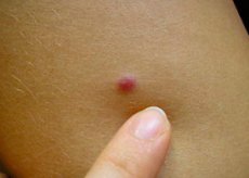
Dangerous symptoms: inflammation of the mole, redness, pain, itching
Last reviewed: 04.07.2025

All iLive content is medically reviewed or fact checked to ensure as much factual accuracy as possible.
We have strict sourcing guidelines and only link to reputable media sites, academic research institutions and, whenever possible, medically peer reviewed studies. Note that the numbers in parentheses ([1], [2], etc.) are clickable links to these studies.
If you feel that any of our content is inaccurate, out-of-date, or otherwise questionable, please select it and press Ctrl + Enter.
Every person notices dark pigmented spots on their body - moles. Some people have single ones, while others have whole scatterings of different marks, differing in size and structure. Sometimes a person is born with a birthmark, and many spots can appear throughout life. These marks can cause certain cosmetic inconveniences, and their changes can be a symptom of dangerous diseases.
What is a mole?
Medical scientists use the term nevus to describe a birthmark. Unlike birthmarks, nevi are not congenital. A birthmark is a small spot that appears during the life cycle on any part of the human body, visually noticeable on the surface of the skin. It consists of the pigment melanin and melanocyte cells. If a birthmark is located in the upper epidermal tissues of the skin, its shape will be flat. The pigmented segment located in the deep dermal tissues will be slightly raised above the skin.
The color spectrum of moles ranges from light coffee to almost black. The color directly depends on the amount of melanin pigment produced by specialized cells (melanocytes).
Flat or convex nevi do not pose a threat if:
- they are uniformly colored, have smooth contours, and are small in size (no more than 5 mm),
- do not tend to increase in area or volume and have not been injured.
Moles that have hair growing from them have a minimal chance of becoming melanoma. In addition to the possibility of malignancy, moles can become inflamed.
Why did the mole become inflamed and what to do? This question often worries people who notice changes that have occurred with the nevus.
Moles are poorly protected from external irritants. There are many factors that provoke inflammation, which in turn leads to pain.
Causes of Mole Inflammation
Factors that contribute to the appearance of inflammation of moles are the following:
- exposure to UV radiation from prolonged exposure to bright sunlight (especially for people with fair skin);
- various injuries (abrasions, cuts).
In these cases, the "gates" open for infection to enter the nevus. Itching, soreness, swelling, redness, and peeling are the symptoms of inflammation. Bloody discharge and intense redness of the skin around the pigmented spot are visual signs of possible transformation of the nevus into a malignant tumor.
Each of the listed symptoms is a reason to consult an oncologist. Confirmation of the presence of melanoma is a good reason for immediate removal of the mole.
Pathogenesis
Moles on the human body can be classified according to various parameters: size, shape, location in skin tissue, etc.
Based on the area they occupy, all nevoid formations are differentiated into:
- Small (up to 15 mm in diameter).
- Medium (size from 15 to 100 mm).
- Large (more than 100 mm in diameter).
- Giant – occupy a certain part of the body (face, chest, arm, etc.).
Small moles are not dangerous and the risk of their transformation into malignant formations is minimal. Malignancy of giant nevi reaches 50%. In the presence of a dangerous neoplasm, it is necessary to be observed by a dermatologist or oncologist. In childhood, the appearance of moles is associated with the migration of pigment cells from deep tissues to the surface of the epidermis.
Depending on the form and origin of nevoid pathology, they are divided into:
- Vascular genesis.
- Hemangiomas are point or volumetric spots on the skin, with the involvement of the vascular wall tissue in the process, having an uneven edge. The color features depend on the type of blood vessel from which they were formed (various shades of pink and red).
- Capillary angiomas. Flat red spots on the surface of the epidermis, causing a cosmetic defect. They pose no danger.
- Cavernous angiomas. Multi-cavity formations of vascular origin, nodular or tuberous in shape, cherry-colored, based in the tissues of the skin. They are dangerous due to the possibility of profuse bleeding in case of injury.
- Nevi of non-vascular genesis. Pigmented skin lesions - from light brown to intense black in color, appearing as various elevations above the skin surface.
Dermatologists diagnose the following types of moles, depending on location, shape, color and size:
- Convex. They are based in deep epidermal tissues and look like a smooth or bumpy formation (no more than 10 mm in diameter) with hair growing in it. The color palette ranges from dark yellow to deep black.
- Flat. Pigmented formations (from light to dark shade), consisting of melanocytes and melanin, localized in the upper layers of the epidermis.
- Lentigo. A large number of pigment spots of a brown or brownish hue. Birthmarks of this type are formed in adolescence or adulthood, turning into a pathological condition - lentigo.
- Congenital melanocytic nevus. Appears as a gray, brown or black spot. Tends to grow as the child gets older.
- Blue nevus. The neoplasm has a hemispherical shape with a diameter of up to 20 mm, rising above the surface of the epidermis. Color - from light blue to dark cornflower blue. Localization sites - limbs, face, buttocks.
- Dysplastic. It is represented by a group of formations of various shapes and a diameter of more than 10 mm, reddish in color with blurred contours.
Moles are divided into melanoma-dangerous and melanoma-safe according to the risk of malignancy. Melanoma-dangerous nevi include: blue, dysplastic and congenital melanocytic.
Symptoms mole inflammation
Symptoms of inflammation of a mole:
- edema;
- redness;
- itching;
- pain in the affected area;
- change in the color of the skin surrounding the mole;
- bloody discharge.
The last two points deserve close attention. They may be a signal of transformation of a benign neoplasm into a malignant one.
 [ 5 ]
[ 5 ]
First signs
The appearance of redness around the nevus signals the initial stage of the inflammatory process. This symptom cannot be ignored. It is necessary to immediately consult a specialist.
It is not difficult to notice the first signs of inflammation of moles on the face, abdomen, and limbs. It is more difficult to diagnose the inflammatory process of nevoid formations on areas of the body that are inaccessible for self-examination. In this case, you will need the help of loved ones.
At the initial stage of inflammation of a mole, the first signs are:
- redness;
- swelling of the site of inflammation;
- pain sensations;
- itching;
- sometimes fever.
To prevent symptoms and manifestations of the inflammatory process in the nevus area, it is necessary to carefully and regularly examine moles. There is a risk of transformation of an inflamed mole into a malignant neoplasm.
[ 6 ]
The mole became inflamed and hurts
People often ask the question: why does a mole become inflamed, what is its cause? The inflammatory process in the nevus area is caused by:
- Mole trauma (careful hygiene, mole rubbing against clothing, insect bites). If you do not pay due attention to the affected mole, the inflammatory reaction can cause deformation of the nevus and provoke its malignancy.
- Intense exposure to ultraviolet radiation. During the daytime in summer, exposed areas of the skin are exposed to intense sunlight. Under their influence, inflammation of the mole may occur, and the person will consider the symptoms to be a manifestation of sunburn.
If inflammation of the skin under the nevoid lesion occurs and discharge forms, then this is a manifestation of the introduction of a bacterial or fungal infection. To determine the pathogen, it is necessary to visit the laboratory and make a smear of the contents from the lesion.
Malignancy of a neoplasm. If a mole becomes inflamed without any apparent cause, this can be interpreted as a sign of its malignancy. In this situation, it is better to visit an oncologist.
The mole became inflamed and reddened
Although moles are benign formations, they cause quite a lot of trouble. The main reason for concern is a change in the structure of the mole, and especially the appearance of a red border.
The primary causes of redness of a nevus or the skin around it are mechanical damage or the beginning of the malignant process.
A mole can be injured by tight or rough clothing, a hard washcloth during hygiene procedures, accidental scratching, etc. In these situations, a reaction of reddening of the mole itself and the skin surrounding it occurs, and sometimes bleeding occurs. Nevi located on the neck, chest, and back are most damaged. In men, moles located in the face and neck area are often injured during shaving. A woman, performing a depilation procedure, risks damaging a mole located in the armpit area, on the legs, or bikini area.
In all the above situations, the mole becomes inflamed, redness and swelling appear. Unreasonable hyperemia of the mole accompanies its malignant transformation.
The redness associated with sunburn will not only affect the nevoid lesion, but will also spread to the entire body part that has been exposed to prolonged aggressive ultraviolet rays.
The appearance of redness and pain in the nevus area is often associated with changes in the hormonal balance in the body (during gestation, taking synthetic hormonal agents).
The mole is inflamed and itchy
For a long time, nevi do not attract attention. But with the appearance of itching in the area of the pigmented lesion, an urgent visit to the oncologist is necessary. One of the reasons for the itching of a nevus is a pathological process associated with uncontrolled cell division, leading to compression of surrounding tissues and structures. During this period, the mole itches intensely, and sometimes changes color, shape and configuration.
Such a dynamic increase in the number of pathological cells may signal the formation of a malignant tumor. Warning signs include: redness around the nevus, itching, and the appearance of liquid discharge from the mole.
Mole became inflamed during pregnancy
The appearance of additional pigment spots during gestation is a natural phenomenon. During this condition, the hormonal balance is transformed. Hormonal imbalances contribute to the intensive appearance and growth of nevi. No pregnant woman is immune from the occurrence of inflammatory phenomena affecting moles. Symptoms of inflammation: redness, swelling, fluid discharge from the nevus, burning and itching are possible. If such signs appear, you should immediately consult a doctor to determine the cause of the inflammation of the mole. After a detailed examination, a specialist will determine what measures should be taken to stop the inflammatory process, and will give recommendations on the proper care of the infected area.
The most common tips for caring for an inflamed mole are:
- avoid exposure to direct UV radiation,
- avoid additional traumatic impact on the nevus,
- do not scratch,
- Squeezing out the contents of a mole is prohibited, even if significant accumulations of exudate are observed.
Removal of inflamed moles during gestation by surgery or laser methods is an extreme measure. Surgical intervention is performed in the only case when there is a high probability of malignancy of the nevus.
Complications and consequences
Factors that provoke adverse effects are divided into two categories: mechanical injuries and ultraviolet radiation.
It is necessary to avoid prolonged exposure to the scorching sun to avoid skin burns and to avoid excessive melanin synthesis. A dangerous consequence is the transformation of a mole into a dangerous malignant neoplasm. In the hot summer period, it is necessary to adhere to the following restrictions:
- during midday hours only short-term exposure to the open sun is permitted;
- visiting solariums is not recommended;
- Beach recreation is permitted only in the morning and evening hours, when UV radiation is less active.
Mechanical damage to a nevus can also have negative consequences. A small scratch can lead to infection, which will cause inflammation of the mole. Chronic traumatic effects of tight clothing, minor cuts to moles when shaving, also pose a risk of infection of the wound, which leads to inflammatory processes in the nevoid focus. Swelling and redness of the skin, accompanied by itching and pain, are observed around the mole. The more often and intensely the nevus is injured, the higher the probability of the onset of a malignant process.
If a mole has changed visually and there are signs of inflammation, you should visit a doctor as soon as possible.
The sudden appearance of inflammation around a mole is one of the threatening symptoms that indicates malignancy of the nevus. Signs of malignancy of a mole are considered to be:
- change the size, color, shape of the edge;
- the appearance of a red border around the nevus;
- increase in the number of nevoid elements.
The danger of inflammation is that with significant mechanical damage to the nevus, bleeding occurs and there is an additional opportunity for the introduction of pathogenic microorganisms.
Diagnostics mole inflammation
Visual diagnosis of moles by a doctor occurs during the examination of the patient. If there is an inflamed nevoid element, the doctor will pay attention to the appearance of a red halo around the nevus, the presence of swelling, and the release of exudate. In order to differentiate infectious inflammation and the onset of a malignant disease, it is necessary to conduct a histological examination.
In case of inflamed moles, a general or biochemical blood test, a general urine test will be of little information. These studies will be required if there is a need to surgically remove the nevus or the doctor suspects a malignant process. If the inflamed nevus has healed within a week, then no additional tests or studies will be required.
Experienced doctors cannot always distinguish an inflamed mole from a malignant neoplasm by visual signs. This serves as a basis for using a biopsy and further histological analysis of the obtained tissue.
In some cases, computer dermatoscopy is used, which allows determining the degeneration of a mole into a malignant oncological disease at an early stage. This diagnostic procedure allows a specialist to study surface changes in detail. There is no traumatic tissue damage during the examination. The doctor analyzes the results and determines the risk of malignancy of the nevus. After conducting detailed and differential diagnostics, the specialist decides on a radical method of treating the mole.
[ 11 ]
How to examine?
Differential diagnosis
Differential diagnostics must be carried out when determining bacterial inflammation of a mole and early stages of skin melanoma. These pathologies have many similar visual (external) symptoms:
- dimensions - 1-3 cm;
- shape - bumpy, nodular, hemispherical;
- surface - shiny, ulcerated, weeping, easily bleeding or crusted;
- color - from pink to black.
Signs of skin melanoma are:
- the presence of "daughter" nodes (satellites);
- pigment inclusions around the tumor;
- enlargement of nearby or peripheral lymph nodes.
- The final diagnosis can only be confirmed by histopathological examination.
Which doctor should I go to if a mole becomes inflamed?
If there is an inflammatory process in a mole, a consultation with a dermatologist or dermato-oncologist who deals with the problems of malignant degeneration of nevi is necessary. Only experienced, highly specialized experts will be able to conduct an accurate diagnosis if a mole is inflamed.
Treatment mole inflammation
If the inflammation of the mole occurred as a result of an injury, the wound surface is small and the nevus is not badly damaged, it will be enough to treat the injury site with an antiseptic solution.
If the inflammation of the mole is chronic (it is located in a place where it is subject to constant trauma), the inflammatory process occurs constantly - it is better to remove such a nevus, without waiting for its possible transformation into a malignant neoplasm. For radical treatment of nevi that are subject to constant trauma, various surgical methods are used.
Medicines
The most well-known way to significantly reduce inflammation is to treat the wound surface with medical ethyl alcohol 70. The procedure is repeated several times a day. To do this, you will need to moisten a sterile cotton swab with alcohol and apply it to the inflamed mole without using force. If there is no medical alcohol in the pharmacy, you can take any alcohol-containing solution.
A simple and easy way to get rid of the inflammatory process is to use streptocide powder. The medicine is available in tablet form. Before use, the tablet should be crushed into powder and sprinkled on the affected area. A few minutes of contact of the drug with the inflammation site will be enough. There is no need to make bandages or fix streptocide powder with a plaster.
Inflamed moles can be treated with an ointment containing an antibiotic, zinc, or phenolic (salicylic) acid.
Folk remedies
Traditional healers have long sought simple and easy ways to quickly get rid of inflammatory processes. Various plants and objects that were available were widely used. Below are several recipes that were used by our predecessors who lived several centuries earlier:
- Lemon and garlic juice. Inflammation around a mole can be relieved by alternately applying garlic and lemon juice. Duration of treatment is 7 days.
- Aloe juice. The juice of this plant has bactericidal properties. It is necessary to squeeze the aloe juice from the leaves 2-3 times a day and treat the inflamed area with it. Only freshly squeezed juice is effective.
- Silver items. In ancient times, the bactericidal properties of silver were used to relieve inflammation. Most often, a silver cross, coin, ring, etc. were used. Any silver item was used to apply to the inflamed area. The only condition was that the item was as clean as possible. The average treatment time is 1 month.
It is necessary to remind that scientists have not yet invented a remedy that will be selectively effective in case of inflammation of a mole. The presence of an infected lesion in the nevus area is a reason to visit a specialist in the nearest future.
Herbal treatment
There are cases when a birthmark has been injured, but seeking medical help is not possible. In such a situation, it is recommended to use herbal remedies.
Compress from celandine tincture. The industrially produced medicine can be bought at a pharmacy without a prescription, or you can make it yourself. In June, collect the leaf part of the plant. Wash the collected leaves thoroughly, then chop finely, fill a 1-liter jar to the top with raw materials, pour 70% medical alcohol and leave to infuse for 14 days. The prepared remedy can be applied to the area of the inflamed mole as a ten-minute compress. It is considered acceptable to treat the inflamed area of the nevus three times with a cotton swab soaked in celandine tincture.
Calendula flower tincture. Tincture from this plant is an excellent anti-inflammatory, antiseptic drug. In addition, it has properties that stimulate the regenerative abilities of body tissues. There is a pharmacy form of calendula tincture, or you can make it yourself. To prepare an alcohol tincture, you need to take 100 ml of vodka and dry calendula flowers (2 tablespoons). Infuse in a dark place for 14 days. Treat the inflamed mole with the prepared product three times a day until completely cured.
Flaxseed oil. To prepare the oil at home, grind flax seeds in a blender until they become flour. Place the resulting mass on gauze and hang it over a container where the oil will drain. The process of separating the oil is quite long, so it will be easier to buy flaxseed oil prepared industrially at a pharmacy or supermarket. To treat an inflamed mole, apply the oil to the affected area at least 4 times a day. It will promote faster healing of the wound.
Homeopathy
Homeopathic treatment for inflammation of a mole is of auxiliary importance, because preparations made from plant materials act last on the skin. Homeopathic preparations have a systemic effect on the body's immune system and thus help reduce inflammation in the lesion. The most commonly used homeopathic remedies are:
- Cutis compositum. Has an anti-inflammatory effect.
- Calendula Salbe Heel. Used to treat long-term non-healing wounds.
- Belladonna Homaccord. Prescribed for inflammatory diseases of various structures of the body.
- Arnica-Heel. Has anti-inflammatory, decongestant, analgesic effect.
These preparations are available in liquid form, packaged in bottles or ampoules for injections of dark glass. When using the preparation orally, 10 drops of liquid dissolved in a small amount of water are usually prescribed. It is allowed to take the medicine directly under the tongue, without diluting it with water. It is advisable to take the homeopathic remedy 20-30 minutes before meals or 1 hour after meals. The duration of the course of treatment and the dosage of the drug are prescribed by the homeopathic doctor for each patient individually.
Surgical treatment
The doctor recommends surgical radical treatment of the mole:
- if the nevus is located in a place that is subject to constant trauma (friction from clothing or accessories, damage from a razor, washcloth, etc.).
- when the contour and color of a mole changes, pain, itching, redness in the area of the nevus appear, which do not decrease for a long time.
Today, doctors can recommend several of the safest methods for removing pigmented benign skin lesions:
- use of liquid nitrogen (cryodestruction);
- application of high voltage electric current (electrocoagulation);
- elimination using laser (laser coagulation).
Prevention
To prevent inflammation and malignant transformation of moles, for prophylactic purposes it is necessary to:
- conduct observation and self-diagnosis of existing moles, for changes in the number of neoplasms, their color, shape, contours;
- seek specialist advice if swelling or redness is detected in the area of the nevus;
- do not expose to traumatic impact;
- avoid prolonged exposure to direct sunlight, use sun protection;
- do not self-medicate suspicious moles;
- If a problem arises or is detected, you must immediately consult a doctor.
Forecast
If the mole has become inflamed due to the introduction of pathogenic bacteria, the prognosis in this case is favorable. But if the inflamed nevus does not heal for a long time, a biopsy or removal of the neoplasm with subsequent histological analysis will be required. If the benign nature of the process is confirmed, a course of therapy aimed at treating the inflammation focus is necessary. If the malignant nature of the process is recorded, then treatment with cytostatic drugs will be required. Treatment started at the initial stage of malignant oncological pathology is very successful and rarely leads to relapses.
[ 16 ]