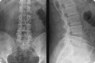
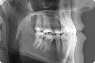
For doctors in traumatology, vertebrology and orthopedics, an X-ray of the lumbosacral spine allows them to diagnose its anatomical abnormalities, injuries and diseases, and then to treat them.
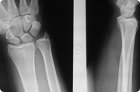
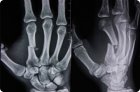
Because X-rays provide an image of the dense structure of bones, X-rays of any joints, including those of the wrist, are the classic method for the initial diagnosis of joint injuries and diseases.
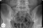
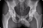
X-ray of the ilio-sacral (ileosacral) joints visualizes the most important anatomical structures of the musculoskeletal system: paired sacroiliac joints that connect the articular surfaces of the sacrum (os sacrum) and the iliac bones (os ilium) entering the pelvic ring.
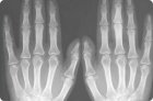
X-rays or X-rays of the fingers - obtaining a fixed black-and-white image of their bones and soft tissues - is a method of radiation diagnostics commonly used in clinical traumatology, orthopedics and surgery.
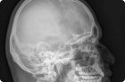
X-ray in medicine is a method of studying the anatomical structures of the body to obtain their projection using x-rays on paper or film, which does not require penetration.
X-ray examination is considered a fairly common diagnostic method, which is used to determine the condition of the musculoskeletal system, detect violations, injuries and diseases.
The most informative methods of instrumental diagnostics of articular and bone pathologies are visualization, and the most accessible of them is an x-ray of the hip joint.
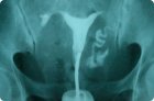
It happens that a woman can not get pregnant. To clarify the cause, it is necessary to conduct an examination, including an x-ray of the fallopian tubes.
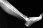
X-ray examination of the lower extremities - X-ray of the leg - fundamental in traumatology and orthopedics and no less important in rheumatology, because it allows doctors to visualize bones and bone structures