Medical expert of the article
New publications
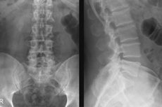
X-ray of the lumbosacral spine
Last reviewed: 06.07.2025

All iLive content is medically reviewed or fact checked to ensure as much factual accuracy as possible.
We have strict sourcing guidelines and only link to reputable media sites, academic research institutions and, whenever possible, medically peer reviewed studies. Note that the numbers in parentheses ([1], [2], etc.) are clickable links to these studies.
If you feel that any of our content is inaccurate, out-of-date, or otherwise questionable, please select it and press Ctrl + Enter.

For doctors of such specializations as traumatology, vertebrology and orthopedics, X-rays of the lumbosacral spine allow them to diagnose its anatomical anomalies, injuries and diseases, and then treat them.
Indications for the procedure
X-ray examination of the lumbosacral - lumbosacral spine is prescribed to patients with pain localized in the area of the L1-L5 and S1-S5 vertebrae, in order to determine their cause and confirm or refute: [ 1 ]
- fracture or other traumatic injury;
- lumbar hyperlordosis;
- intervertebral hernia;
- arthritis and osteoarthritis;
- osteoarthrosis or osteochondrosis;
- displacement of lumbar vertebrae (spondylolisthesis);
- spondylitis;
- sclerotic and degenerative changes in the vertebrae - lumbar spondylosis;
- dysplasia/hypoplasia of the articular processes of the vertebrae;
- ankylosing spondylitis (Bechterew's disease);
- deforming spondyloarthrosis (pathology of the facet joints);
- ossification of the ligaments of the spine (idiopathic lumbar hyperostosis),
- scoliosis;
- sacralization or lumbarization of the lumbar and sacral vertebrae.
X-rays are used to monitor the progression of diseases or determine the effectiveness of their treatment, as well as after surgery. [ 2 ]
X-ray of the sacroiliac joints - two sacroiliac joints connecting the sacrum (os sacrum), located below the lumbar region, with the iliac bones (ossis ilium) of the pelvis, that is, X-ray of the iliosacral joints of the sacral spine - allows you to determine the cause of pain and stiffness of movement, including: arthrosis and arthritis; inflammatory process (sacroiliitis); degenerative-dystrophic changes in bone structures in osteoporosis. And also to differentiate neurogenic, muscular or somatic pain in the sacrum from vertebrogenic pain syndrome.
Preparation
X-rays of these segments of the spinal column require preparation. Firstly, three days before the examination it is recommended to stop eating foods that cause flatulence (increased gas formation in the intestines).
Secondly, an enema is done before an X-ray of the lumbosacral spine: bowel cleansing is necessary to obtain better quality images.
Directly in the X-ray room, the patient must remove everything made of metal.
Part of the abdominal region, the mediastinal region, and the thyroid gland are protected with lead pads.
Technique X-rays of the lumbosacral spine.
Standard images of the lumbosacral region and ileosacral joints are taken in direct and lateral projections. An image at an angle (in an oblique projection) may be required separately.
The patient's position for obtaining a frontal (anterior-posterior) image is lying on his back or stomach (depending on the requirements of the attending physician); for a lateral image, lying on his side. [ 3 ]
In addition, to assess the stability of the spine under physiological stress, a functional X-ray of the lumbosacral spine is performed: images are taken in the lateral projection with the patient standing, sitting, and leaning forward.
More details in the publication - X-ray of the lumbar region with functional tests
What does an x-ray of the sacral spine show?
In osteoarthritis and osteochondrosis, an X-ray of the sacral spine shows a decrease in the width of the intervertebral space - the result is a reduction in the height of the intervertebral disc; displacement and deformation of the processes of the vertebral bodies and the vertebrae themselves; bone growths (osteophytes) are observed on the sides of the vertebrae.
More details in the materials:
- X-ray signs of osteochondrosis
- Diagnosis of osteochondrosis of the lumbosacral spine
- X-ray signs of bone and joint diseases
In ankylosing spondylitis, the image shows symmetrical changes in the sacroiliac joint: elements of ligament calcification, vertically protruding osteophytes (syndesmophytes). [ 4 ]
The presence of an inflammatory process in the iliosacral joints (sacroiliitis) is indicated by the widening of the joint space visualized in the image, the absence of clear contours of the endplates of the vertebrae and the proliferation of their bone tissue.
The radiograph shows the bone fusion of the last lumbar vertebra (L5) and the first sacral vertebra (S1). The condition of the L5 vertebra and the lack of fusion of its arch (spondylolysis) is shown in the oblique projection image.
Complications after the procedure
There can be no consequences after a single X-ray procedure (with a radiation dose of 0.7 mSv). There are also no complications after the procedure.
It should be borne in mind that each x-ray performed (regardless of which organ is examined) is recorded in the patient's medical record, and the indicator of the total dose of ionizing radiation received over 12 months should not be higher than 1 mSv. So the risks associated with radiation may be associated with exceeding this indicator.

