Medical expert of the article
New publications
The sacroiliac joint.
Last reviewed: 07.07.2025

All iLive content is medically reviewed or fact checked to ensure as much factual accuracy as possible.
We have strict sourcing guidelines and only link to reputable media sites, academic research institutions and, whenever possible, medically peer reviewed studies. Note that the numbers in parentheses ([1], [2], etc.) are clickable links to these studies.
If you feel that any of our content is inaccurate, out-of-date, or otherwise questionable, please select it and press Ctrl + Enter.
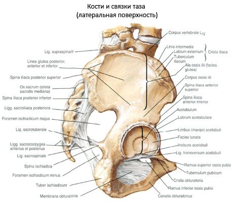
The sacroiliac joint (art. sacroiliaca) is formed by the ear-shaped surfaces of the pelvic bone and sacrum. The joint capsule is thick, tightly stretched, attached along the edges of the articular surfaces, merging with the periosteum of the pelvic bone and sacrum.
The ligaments that strengthen the joint are thick and strong. The ventral (anterior) sacroiliac ligaments (ligg. sacroiliaca anteriora) connect the anterior edges of the articulating surfaces. The posterior side of the capsule is reinforced by the dorsal (posterior) sacroiliac ligaments (ligg. sacroiliaca posteriora). The strongest are the interosseous sacroiliac ligaments (ligg. sacroiliaa interossea), located on the posterior surface of the joint and connecting both articulating bones. The also present iliolumbar ligament (lig. iliolumbale) connects the transverse processes of the IV and V lumbar vertebrae with the tuberosity of the ilium. The sacroiliac joint is flat in shape of the articular surfaces. However, movements in it are practically impossible. This is due to the complex relief of the articulating surfaces, tightly stretched joint capsule and ligaments.

 [ 1 ]
[ 1 ]
Where does it hurt?
What do need to examine?
How to examine?
What tests are needed?

