Medical expert of the article

New publications
Head X-ray
Last reviewed: 04.07.2025

All iLive content is medically reviewed or fact checked to ensure as much factual accuracy as possible.
We have strict sourcing guidelines and only link to reputable media sites, academic research institutions and, whenever possible, medically peer reviewed studies. Note that the numbers in parentheses ([1], [2], etc.) are clickable links to these studies.
If you feel that any of our content is inaccurate, out-of-date, or otherwise questionable, please select it and press Ctrl + Enter.
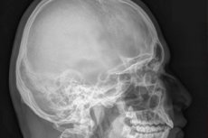
The most accessible and quite informative method of visualizing the bones of the skull is an X-ray of the head or craniography. This study is usually prescribed when there is a suspicion of pathologies of bone structures, however, even from a general X-ray image it is possible to assume the presence of a brain tumor, hematoma or ischemic area, even intracranial hypertension, after which a search can be conducted in a specific direction.
Craniography has been used for diagnostic purposes for decades and has not lost its relevance.
Indications for the procedure
X-rays of the skull bones are always indicated in patients with head injuries. [ 1 ]
The basis for conducting such a study may be a suspicion of congenital and acquired pathologies of the cranium - visible violation of symmetry, size and shape, patient complaints of tremors of the limbs, impaired coordination of movements, frequent and excruciating headaches, dizziness, nausea, deterioration of vision and hearing, pain when moving the maxillofacial structures.
Technique head X-rays
The head X-ray is taken depending on the required angle and the equipment used in a sitting or lying position, sometimes standing. The patient must remain motionless for several minutes during the imaging, which the radiologist warns him about. Foam pads, pillows, and fixing belts can be used to ensure comfort when holding the head in the desired position. Lead vests and aprons are used to protect body parts that are not subject to examination.
A child's head X-ray is done only for vital indications. In childhood, doctors try to use alternative and safer imaging methods, such as ultrasound or MRI. However, the condition of bone structures can be best assessed by an X-ray. Therefore, if a child hits his head, it is better to exclude the possibility of damage to the skull bones.
An X-ray of the head of a child under one year old is also done in case of head injuries, including those received during childbirth, as well as in case of suspected congenital pathologies, since without diagnosis the time for effective treatment can be missed.
Children are carefully screened for body parts that are not subject to examination. The most difficult thing when taking an X-ray of a child is to ensure that he or she remains motionless. The smallest children are usually given a head X-ray under sedation; older children are persuaded, calmed, and fixed in the desired position. For this, parents are called upon to help. [ 2 ]
Pregnancy is a contraindication for X-ray examination. However, there are circumstances (blows, falls, traffic accidents) when X-ray of the head during pregnancy is necessary. In this case, the body and especially the abdomen are covered with capes that do not allow X-rays to pass through.
Contraindications to the procedure
Absolute contraindications to routine examination using radiation methods are:
- the presence of a mental illness that makes it impossible for the patient to adequately perceive the requirements for the procedure - he does not understand the need to sit or stand in a certain way, remain motionless for a short time, etc.;
- Also, the examination is prohibited for pregnant women and children under 15 years of age, since radiation can have a teratogenic effect and negatively affect the development of bones in the child.
In emergency cases, when an X-ray of the head is necessary for vital indications, it is performed on all categories of patients, carefully observing preventive measures, immobilizing with medication people who are unable to remain motionless.
X-ray examination is not performed on people with metal or electronic implants in the diagnostic area. [ 3 ]
A temporary recommendation is to postpone the planned procedure until a more favorable period for people with a reduced immune status.
Is a head x-ray harmful?
The diagnostic procedure is practically harmless, the radiation dose is low and the exposure time is very short. Even several X-ray examinations of the skull bones per year will not cause significant harm. On average, the radiation dose during an X-ray of the head is 0.12 mSv. For comparison, epidemiological studies on people indicate that the minimum oncologically dangerous radiation dose received in childhood begins with 50 mSv. This same indicator is on average more than 100 mSv.
The sanitary norm is considered to be the radiation dose received during X-ray examinations, 1 mSv or six to seven roentgens annually. Therefore, even if in one year you had to undergo, for example, eight procedures of radiological diagnostics, then in the next – there may not be a single one.
And if you compare the danger of radiation during a head x-ray with the danger of losing your life or becoming disabled, then you can exceed the norm written in reference books, since an accurate diagnosis increases the guarantee of successful treatment.
Normal performance
Based on the patient's complaints, anamnesis and clinical manifestations, an X-ray examination of the skull bones in one or more projections may be prescribed. Sometimes a targeted examination of a specific area of the head is prescribed.
In case of trauma, congenital pathologies, patient complaints of headache, dizziness, and impaired coordination, a survey X-ray of the skull is performed. This reveals fractures and cracks in the bones, displacement of bone fragments; developmental anomalies; curvature of the nasal septum and diseases of the paranasal sinuses.
In addition, an X-ray may indicate osteomyelitis of the cranial bones by the presence of calcification foci (white areas impermeable to rays), and osteoporosis by areas of bone rarefaction. Intracranial calcification foci are interpreted as signs of chronic subdural hemorrhage; oligodendromas and meningiomas (tumor calcification) look approximately the same, only with a more distinct rounded shape. [ 4 ]
The X-ray can also show vascular changes characteristic of high intracranial pressure, abnormalities specific to the metabolic disorder associated with excessive secretion of growth hormone (acromegaly), and softening of the bones associated with Paget's disease. A single X-ray may not always be sufficient to make a definitive diagnosis, but it may indicate the direction of subsequent diagnostic investigation.
Quite often, people are prescribed targeted x-rays of the sella turcica area to detect prolactinoma, to clarify the presence of osteoporosis, and to better examine the features of the vascular pattern if intracranial hypertension is suspected.
A popular study is an X-ray of the temporomandibular joints, which shows arthritis or arthrosis of the joint of the same name, and its dysfunction. Such a picture is taken in two positions: in one, the patient's mouth is open, in the other, it is closed.
In case of purulent mastoiditis, an X-ray of the temporal bone area is prescribed; targeted X-ray of the zygomatic bone can determine the cause of pain when chewing and other jaw movements.
In traumatic brain injuries, fractures in the eye socket area are often encountered. This examination can also detect the presence of a foreign body in the eye. [ 5 ]
The nasal bones, which are often the most prominent part of the face due to facial injuries, are specifically illuminated. A popular appointment is radiography of the mandibular region. They are mainly prescribed when fractures are suspected, but tumors and some inflammatory diseases can be detected in this way.
Complications after the procedure
When X-raying any area of the body, the impact of low-intensity sources of ionizing radiation occurs directly at the time of the procedure. Electromagnetic waves used in X-ray equipment do not accumulate in the body. Therefore, there is nothing to "remove" from the body after the procedure. Even when repeating X-rays of the head, no immediate complications can arise after the procedure. Therefore, when people complain that they feel bad after an X-ray of the head, this is explained by other factors. Firstly, they are unlikely to have felt well before the examination, there must have been some complaints, since X-ray diagnostics are not done just like that, on a whim. Secondly, suspiciousness, anxiety, and the expectation of complications also do their job.
However, it is recommended to do head X-ray only on doctor's prescription, in addition, if it is not a one-time procedure, it is advisable to monitor the radiation dose received during diagnostic procedures during life. Because the main consequence after the procedure is exceeding the permissible average annual radiation dose, but for this you need to undergo more than twenty examinations per year. So there is no need to be afraid of complications.
However, refusing to undergo diagnostics can lead to serious consequences associated with a risk to life.
Reviews of head x-rays are the most favorable. The procedure is short, does not cause any preliminary troubles and does not cause unpleasant sensations. Advice on increasing the effectiveness of the examination and reducing the radiation dose - if possible, choose a room equipped with a digital x-ray machine.
It happens, of course, that after an X-ray there is a need for a computed tomography scan (if the patient has a high bone density, a layer-by-layer study is more informative) or a magnetic resonance imaging study (when the presence of vascular pathologies or brain tissue is suspected).
For the study of damage to bone structures, radiography remains the method of choice due to its low cost and the availability of X-ray rooms in almost all outpatient departments.