Medical expert of the article

New publications
Lumbar X-ray with functional tests: how to prepare and how is it done?
Last reviewed: 03.07.2025

All iLive content is medically reviewed or fact checked to ensure as much factual accuracy as possible.
We have strict sourcing guidelines and only link to reputable media sites, academic research institutions and, whenever possible, medically peer reviewed studies. Note that the numbers in parentheses ([1], [2], etc.) are clickable links to these studies.
If you feel that any of our content is inaccurate, out-of-date, or otherwise questionable, please select it and press Ctrl + Enter.
The most accessible diagnostics that allows visualizing the internal structure and assessing the condition of the skeletal bones of the spine is radiography. The procedure is painless, does not require complex preparation, is affordable and is performed quickly in almost all medical institutions. X-rays of the lumbar spine are used primarily to identify defects in bone structures (fractures, osteophytes, curvatures, displacements, shifts, neoplasms and inflammatory processes), since their tissue is the most contrasting and clearly visible on the radiograph.
This examination alone is sometimes insufficient to assess the condition of the fibrocartilaginous structures of the intervertebral discs and soft tissues. To accurately establish that there are muscle and ligament strains, nerve and vascular damage, or spinal hernias, it is necessary to additionally use other methods.
Indications for the procedure
A patient can be referred for an X-ray of the lumbar spine by various specialists: a therapist, a neurologist, a vertebrologist, an oncologist, an orthopedist. When referring a patient to the diagnostic department, the doctor specifies the area of the spinal column to be examined and the presumed diagnosis.
The basis for referring a patient for an X-ray may be:
- complaints of numbness and muscle cramps in the lower extremities;
- assumptions about possible deformations of the spinal column in this area: displacement of discs, hernias, bone growths, compressions causing pain, tingling, numbness in the lumbar region;
- traumatic injuries: fractures, dislocations, subluxations of the lumbar spine;
- oncological diseases of this localization, primary and metastatic;
- collagenoses;
- suspected infection of the spinal column – osteomyelitis, syphilis, tuberculosis;
- suspicion of congenital anomalies of the spine;
- pre- and postoperative examination;
- monitoring the results of the therapy.
 [
[ Preparation
Before this diagnostic procedure, it is necessary to carry out simple preparation for the examination. The procedure of X-raying the lower part of the spinal column is performed in the first half of the day on an empty stomach. Beforehand, the evening before, it is necessary to cleanse the intestines. This is done so that the gases accumulated in the intestines do not distort the clarity of the image and, accordingly, the result of the examination.
It seems to be simple and clear, however, how to cleanse the intestines before an X-ray examination? There are several ways to cleanse.
You can seek medical help and undergo colon hydrotherapy. The procedure of cleaning the intestines with a stream of water will take about twenty minutes, will cost a certain amount of money and time to visit a medical institution by appointment.
Most patients prefer not to bother and do a cleansing enema at home before an X-ray. The equipment for this procedure is usually available in everyone's medicine cabinet - a silicone (rubber) bulb or Esmarch's mug. Using any of these devices, 1.5-2 liters of solution at a temperature of about 37-38℃ should be poured into the intestines of an adult. The simplest and most popular solution is boiled water with salt dissolved in it in the following proportions: a teaspoon of salt per ½ liter of water, therefore, three spoons are dissolved in 1.5 liters, and four in two.
You can brew chamomile from a pharmacy according to the recipe provided in the instructions on the package. A cleansing enema before an X-ray with such an infusion removes gases well and sanitizes the intestines.
When the solution is ready and poured into Esmarch's mug, the patient lies on his left side, slightly bends his knees and inserts the tip, previously lubricated with Vaseline, into the rectum, squeezes the hose and slowly pours the solution into the intestine. After that, you need to try to keep the solution inside for as long as possible. Then - visit the toilet and get rid of the contents of the intestine.
For those who cannot stand the enema cleansing procedure, other methods can be used: drink several (5-7) glasses of salted water at night or use a special medicine, Fortrans, a laxative designed to cleanse the intestines before operations and diagnostic procedures. The drug is diluted with water in proportions of one packet per liter, and the amount of water is calculated from the ratio of 1 liter per 15-20 kg of body weight. Drink the laxative solution slowly. The absorption rate of the solution should be about a liter per hour. The entire portion of the solution can be drunk at night in one sitting, or divided into two portions.
There are different options. They are clearly described in the instructions for the laxative. The patient chooses the most acceptable option for himself depending on the time of the procedure and his own ability to absorb a large amount of liquid. Fortrans is an effective and fairly safe drug, but, like all medications, there are contraindications to its use. These are sensitization, damage to the intestinal mucosa for various reasons - inflammation, tumors, polyposis, the risk of obstruction, by the way, an enema is also undesirable in these cases. If you are taking medications, you should familiarize yourself with the features of interactions in the instructions for Fortrans.
It should also be noted that enemas are not given and laxatives are not taken immediately after meals. It is necessary that at least three hours have passed since your last meal, and this will be your last meal before the procedure. You can drink clean, non-carbonated water in the evening if you want, but not liters, but a few sips.
Diet before an X-ray of the lumbar spine is also important. About two to three days before the expected diagnostic procedure, you should not eat hard-to-digest foods and foods that promote gas formation. These include fresh buns, cookies and other sweets, carbonated drinks, coffee and alcohol, fresh milk, bread, legumes, cabbage in any form, smoked meat and fish, sausages, raw vegetables and fruits.
Many people wonder what they can eat before a lumbar X-ray. Relax, the diet is not strict. If you eat a piece of bread (not half a loaf) with a stew or a bowl of soup, the cleansing event will level it all out. The same applies to a glass of milk and a sandwich with sausage. However, if there is an opportunity to choose, then preference should be given to easily digestible foods: soups, vegetable purees, meat broths. You can have them with meat or fish, but boiled or baked. Of the drinks, it is better to give preference to tea and plain clean water. Of course, you can do without soda, alcohol, coffee and pea soup for three or four days. Cabbage also causes increased gas formation in many people. In general, everyone knows their reaction to foods and can predict the result. If there are problems with digestion, during the preparation period, you can take a tablet of an enzyme preparation (mezim, festal) before eating, which promotes digestion. Flatulence that appears at an inopportune time can also be dealt with using activated charcoal or enterosgel.
On the day before the X-ray, it is recommended to have your last meal no later than six o'clock, so that the peak of cleansing activities does not occur late at night.
In addition, when putting on underwear in the morning before the procedure, make sure that it does not have any decorations - metal coating, rhinestones.
Technique lumbar spine X-rays.
On the day of the examination, it is forbidden to eat, drink, or smoke. In the X-ray room, before taking the place indicated by the doctor and assuming the required position, the patient must remove all metal items from his body except for outerwear (he will undress to his underwear).
Lumbar spine x-rays are performed in most cases with the patient lying on his back (direct posterior projection) or on his side (lateral), sometimes, depending on the capabilities of the equipment, sitting or standing. If additional information is needed, an oblique spine examination may be prescribed.
The patient lies down on the table and takes the necessary position, the areas of the body adjacent to the examined (neck and chest) are covered with a lead vest to protect the organs located there from radiation. During the imaging, the patient should not move or even breathe, carefully following the instructions of the radiologist. The procedure itself takes a couple of minutes, the images are usually ready in a quarter of an hour.
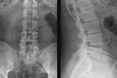
Based on the diagnostic version of the images already obtained, as well as taking into account the patient's complaints, an X-ray of the lumbar spine with functional tests may be prescribed. The purpose of such a study is to assess the mobility of this section. The image is taken in the maximum positions of the bent and straightened spine. Images taken in a standing or sitting position are considered preferable and most informative, but depending on the type of equipment, the objectives of the examination and the patient's health, a lying position may also be used.
Functional X-ray of the lumbar spine is done in lateral projection.
- The patient stands, touching the vertical X-ray stand with his side. First, he leans forward as much as possible, trying to touch the floor with his fingers, with his knees straightened – this is the position in which the first image is taken. Then, straightening up, he bends back as much as possible, raising his arms upward, clasping his palms together at the back of his head – the position of the second image.
- The patient sits, pressing the side of the body to the stand, leaning forward, clasping the knees with crossed arms, and resting the elbows on the hips (first picture). From the same sitting position, to take the second picture, the patient bends back as much as possible, throwing back the head and arching the sternum.
- The flexion shot is taken lying on the side in the fetal position, with the head resting on the bent arm. The second shot (maximum extension) is taken standing at the counter, with the patient simply holding onto the edge of the table with one hand.
Contraindications to the procedure
X-rays are not performed on patients with metal or electronic implants.
Patients with mental illnesses that make it impossible to undergo the procedure due to a lack of understanding of the need to comply with certain requirements (take the necessary position, hold their breath) are not given routine radiography.
Pregnancy and childhood under 15 years of age are absolute contraindications to routine radiography due to the risk of undesirable effects on the intrauterine development of the future child and bone growth in childhood. The exception is cases when this diagnostics can save the life of this category of the population - acute injuries and the need for urgent surgical intervention. Emergency radiography for vital indications is carried out for almost all categories of the population, observing safety precautions.
X-rays may be temporarily contraindicated if the patient is unable to remain still even for a short time; the procedure may be cancelled due to inappropriateness due to lack of bowel preparation.
For people with weakened immunity, routine diagnostics may be postponed until a more favorable period.
In overweight people, this test may not be very informative because layers of subcutaneous fat make the X-ray images less clear.
Normal performance
X-ray images can show changes in the structure of the bone tissue of the vertebrae (fracture, crack, wear, thinning and deformation of bones and cartilage tissue), various pathologies of the spinal column (curvature, narrowing, displacement of vertebrae, cysts, inflammatory processes), suggest the presence of neoplasms, hernias and deformations of the shock-absorbing discs between the vertebrae.
X-rays can diagnose lumbosacral osteochondrosis, osteoporosis, radiculitis, spondylolisthesis, fracture, disc rupture, bone spurs and other growths. Sometimes pathological processes unrelated to spinal diseases are diagnosed as accidental findings.
Individually, after taking pictures in the required projections and having certain complaints, the doctor may prescribe additional examinations. An X-ray of the lumbar spine with functional tests allows you to evaluate the full range of motion in this area. This examination is necessary when the patient has a partial or complete limitation of motor skills of body parts associated with pathologies of this localization.
Description of X-ray of the lumbar spine
In a normal image, the visible area of the lower spine should be smooth, without curvatures, the number of vertebrae, their shape and size should also correspond to the required parameters, the integrity of the bone tissue structures should not be damaged, and the soft tissues surrounding the spine should be without swelling.
X-rays reveal signs of major spinal diseases or suggest trouble and indicate the localization of the pathological process.
The image is made in black and white with areas of different intensity. Bones are the lightest, almost white, clear structures, and soft tissues are very poorly visible, since X-rays pass through them almost completely. Fractures are clearly visible in the image - they look like dark uneven gaps (cracks) crossing the bone, at the fracture sites there may be displacements - misalignment of the lines of the lateral edges of the bone. Scoliosis is visualized in the image as an asymmetrical arrangement of the spinal column (its deviation to any side).
Lumbar osteochondrosis is diagnosed by such signs as a decrease in the clearance of the gap between the vertebrae, in which the intervertebral disc is located. Due to upright walking, the load on the lower parts of the spine is the highest in humans; they are forced to constantly cushion it when running, jumping, walking. It is at the level of the lumbar region that pathological changes are detected first.
The presence of osteochondrosis is also indicated by the presence of compensatory degenerative changes in the form of osteophytes (marginal growths on the vertebral body) that form in places of constant (chronic) damage to the spinal ligaments.
A complication of osteochondrosis, spondylosis, is visualized in the image as beak-shaped growths connecting adjacent vertebrae.
A decrease in the density of the bone tissue of the vertebrae is also noticeable; in areas of reduced density, the film is more strongly exposed, and these areas become darker (gray, not white).
The stages of the disease can also be determined by an X-ray: the first stage corresponds to a decrease in the intervertebral space by a height of no more than a third of the height of the vertebra; the second – up to half. The third stage corresponds to the remaining size of the intervertebral space, which is no more than a third of the height of the vertebral body.
On X-ray, you can see lumbarization of the first sacral vertebra (S1). This pathology looks like an additional fragment of the spine, separated from the main axis by enlightenment. A congenital defect, usually detected accidentally on X-ray. It is one of the causes of lumbago, scoliosis and early osteochondrosis. In this case, the first sacral vertebra is separated from the sacrum partially or completely, becoming the sixth lumbar (normally there are five).
Sacralization of the fifth lumbar vertebra (L5) shows that there are not enough lumbar vertebrae. There are four of them instead of the required five and there are no facet joints, which also leads to early osteochondrosis caused by increased load on the remaining ones due to the absence of the vertebra.
A lumbar disc herniation is determined by the presence of some indirect signs. Its existence can be suspected rather than accurately determined. When a hernia occurs, soft tissues are affected, so it is not visible on an X-ray. Frontal and lateral images are analyzed and compared. It is possible to assume that a hernia may occur if the vertebrae look like high-intensity large rectangular formations with slightly concave sides and rounded corners. The lateral projection shows the presence of osteophytes. Experienced radiologists analyze the contours of the vertebrae, the distances between them, the intensity of the rays passing through the bone tissue, but an error is still possible - such signs may indicate dislocations and fractures, neoplasms, scoliosis and kyphosis.
Sometimes, X-rays show dark spots with clear rounded contours, indicating the possible presence of neoplasms (not necessarily the spine, others located in this area can also be projected). Darker areas with blurred, unclear boundaries can be interpreted as swelling of inflamed tissues. However, no doctor can say exactly what dark spots on X-rays in the lumbar region mean. Additional studies are needed.
Complications after the procedure
In medicine, low-intensity X-ray irradiation is used for diagnostic purposes, which has a short-term effect. Even if this diagnostic procedure is repeated several times, no immediate consequences can arise after the procedure. And the long-term risks, for example, of developing oncopathology in the future in people who have undergone this examination and those who have never undergone it are practically the same.
High-energy electromagnetic waves used in X-ray equipment are not capable of accumulating in body tissues like radioactive chemical elements, so no measures to remove radiation are required after an X-ray examination.
However, you should undergo X-ray examination only as prescribed by a doctor and monitor the radiation dose received during your life. The diagnostic examination is carried out without penetrating the body, it is painless and does not require post-procedure care. In our case, the radiation dose on conventional equipment is 0.7 mSv, on digital equipment - 0.08 mSv, received per procedure (for comparison: the permissible annual dose for radiologists should not exceed 20-50 mSv).
The main complication after the procedure is exceeding the permissible radiation threshold, and to exceed it even on old equipment, you need to take an X-ray of the lumbar spine more than twenty times a year.
So, we have found out that complications do not arise after the X-ray procedure. But refusal of diagnostics can lead to serious consequences, it is possible to "overlook" fractures, dislocations, neoplasms, degenerative changes. Inadequate treatment leads to complications of diseases, constant pain and discomfort, loss of the ability to walk independently.
Reviews of the procedure are the most favorable, it is short-term, does not cause any unpleasant sensations and the patient's condition before and after the procedure does not change. The main impressions and questions concern preparation for X-rays, in particular, bowel cleansing. Very good reviews of Fortrans. Patients with experience, who have something to compare, recommend not doing an enema, but cleansing the intestines with this product.
Due to the low cost of X-ray, the presence of X-ray laboratories in almost all outpatient departments, and its high information content, it is very common. Another piece of advice given by "seasoned" patients: if possible, take X-rays in rooms equipped with the most modern equipment. It has many advantages - from the comfort of the patient and a higher-quality image to the lowest radiation doses.
Analogs to X-ray
An alternative radiation diagnostic method is computed tomography (CT). Unlike an X-ray, the doctor receives a clearer three-dimensional image that can be copied to a laser disk or flash drive, or sent by e-mail. CT can be done for patients with implants. However, this type of examination gives the most significant radiation exposure. The radiation dose for CT of the lumbar spine is 5.4 mSv.
Magnetic resonance imaging (MRI) is quite informative and is considered safe. It can also be used to diagnose injuries and diseases of the spine. However, in addition to the high cost of the study, radiation methods (X-ray and computed tomography) are considered more informative when examining the bone structures of the spine. MRI better visualizes soft tissues, vessels and nerves, as well as neoplasms in them.
When examining a patient, various methods can be used, the choice depends on the patient's complaints and preliminary diagnosis, but an X-ray of the lumbar spine is quite universal and informative, and, importantly, accessible. In some cases, it can give a complete picture. If necessary, it can be supplemented with MRI.