Medical expert of the article

New publications
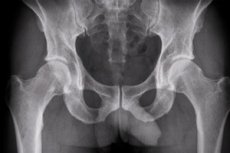
Hip X-ray in two projections
Last reviewed: 03.07.2025

All iLive content is medically reviewed or fact checked to ensure as much factual accuracy as possible.
We have strict sourcing guidelines and only link to reputable media sites, academic research institutions and, whenever possible, medically peer reviewed studies. Note that the numbers in parentheses ([1], [2], etc.) are clickable links to these studies.
If you feel that any of our content is inaccurate, out-of-date, or otherwise questionable, please select it and press Ctrl + Enter.
The most informative methods of instrumental diagnostics of joint and bone pathologies are visualization, and the most accessible of them is X-ray of the hip joint.
Plain radiographic examination is a fundamental approach to diagnosis and treatment decision-making of the hip joint. [ 1 ]
Indications for the procedure
By referring a patient for an X-ray, a traumatologist, orthopedist, surgeon or rheumatologist has the opportunity to assess the condition of the structures of a given bone joint.
The most common indications for X-ray diagnostics of the hip joints concern:
- traumatic injuries to the hip area, in particular, fractures of the femoral neck;
- congenital dislocation or dysplasia of the hip joints;
- juvenile epiphysiolitis of the femoral head;
- arthritis, arthrosis of the hip joint, deforming arthrosis or coxarthrosis;
- coxitis (inflammation of the hip joint);
- necrosis of the femoral head;
- osteoarthritis, osteomyelitis and osteochondromatosis;
- articular cysts and other formations;
- bone tuberculosis affecting the joints.
In principle, patient complaints about perceived pain in the hip joint are considered sufficient reason to prescribe an X-ray - to establish their exact cause. If the above diseases and conditions are absent, the protocol (description) of the X-ray image will indicate that the X-ray is normal. This means that the obtained images of all joint elements do not have anatomical abnormalities, for more details see - Hip joint
X-rays of the hip joints in children are performed according to strict indications - only after the child reaches nine months. The main pathology is congenital hip dislocation. In addition, X-rays can be prescribed for pain in the hip joint in children of different ages.
Preparation
Preparation for an X-ray involves limiting the intake of foods that cause flatulence and cleansing the intestines (using an enema) several hours before visiting the X-ray room.
Immediately before the procedure, the patient is given protection in the form of special shielding coatings that prevent the penetration of X-ray radiation into other parts of the body and internal organs.
Technique hip x-rays.
The standardized technique of performing radiography depends little on the method used - analog or digital. If in the first case the procedure lasts about 10 minutes, and the image is obtained on film, then with the second method the time is cut in half, and the image can be in two formats, including digital.
The maximum visual information is provided by an X-ray of the hip joint in two projections: in a direct projection (or frontal), obtained by focusing the X-ray tube perpendicular to the plane of the body - from the front or back, and axial (transverse or horizontal plane), fixing the elements of the joint from top to bottom - along the femur. The image can also be taken with a lateral projection, that is, the patient must lie on his side, bending the leg at the knee and hip joints.
Conventional radiography usually involves taking anteroposterior and lateral radiographs of the hip. The anteroposterior radiograph of the hip includes images of both sides of the hip on the same film and extends toward the midpoint of the line joining the upper part of the pubic symphysis and the anterior superior iliac spine; the distance between the x-ray tube and the film should be 1.2 meters. When anteroposterior radiographs of the hip are taken in the supine position, one of the most common errors is distortion of the image due to external rotation of the hip.
Thus, either both patellas must be pointed forward or the lower limbs must be internally rotated 15-20° to accommodate femoral antiversion on anteroposterior hip radiographs.
If an X-ray of the hip joints is performed according to Lauenstein (Lauenstein), the patient's position is as follows: lying on the back, one leg is bent at the knee (at an angle of 30, 45 or 90°), while its foot rests on the shin of the straightened leg; the thigh of the bent limb is maximally abducted to the side so that the hip joint takes a position of external rotation (that is, the head of the femur rotates in the acetabulum).
It should be borne in mind that X-rays of the pelvic bones and hip joints in young children do not show the exact outlines of the joint structures, since their main tissue is cartilage, which X-rays do not display. Therefore, the interpretation of the resulting X-ray - with the determination of the displacement of the femoral head in relation to the acetabulum of the pelvic bone - is carried out by superimposing a special grid on the image, the main and auxiliary lines of which correspond to the anatomically normal location of the joint structures. Using these lines, various parameters are measured, including the center of the acetabulum and the degree of inclination of its roof (acetabular angle), the angle of forward deviation of the femoral neck, etc.
And if there is a suspicion of a violation of the development of ossification nuclei (especially in premature babies), an ultrasound examination of the hip joint should be done on a one and a half to two month old child.
Contraindications to the procedure
X-rays are not performed in cases of any acute infections, elevated temperature, bleeding, or inflammation of the synovial bursa of the joint and periarticular muscle tendons.
It is strictly contraindicated to perform X-rays on pregnant women and children in the first nine months of life (although in pediatrics the ban on X-rays applies up to the age of 14).
Normal performance
Each radiograph provides important information needed to accurately diagnose hip disorders [ 2 ]. Typically, the anteroposterior profile provides information about the shape of the acetabulum, while other lateral images provide information about the proximal femur, including the femoral head.
The following information can be obtained from radiographs of the anteroposterior hip:
- leg length,
- neck angle,
- acetabular coverage: lateral central rim (CE) inclination angle and femoral head extrusion index,
- acetabulum depth,
- acetabular tilt,
- acetabulum version,
- sphericity of the head and
- width of the joint space.
Lateral radiographs of the hip joints evaluate the shape and displacement of the articulation of the femoral head and femur, as well as the displacement of the alpha angle. [ 3 ]
Taking into account the Idelberg-Frank angle, the Wiberg angle and the MZ-distance of decentralization, statistical methods can distinguish normal and pathological joints: in adults, hip joint values from 6 to 15 indicate a normal joint shape; values between 16 and 21 indicate a slight deformation, and values from 22 and above indicate a serious deformation; in children, values from 15 and above are pathological. [ 4 ]
It is important to emphasize the need to use radiographs in conjunction with physical examination results, as imaging findings are not always associated with the presence of pain, and vice versa.[ 5 ]
X-ray signs of hip dislocation
On X-ray, signs of hip dislocation or dysplasia are visualized as vertical and lateral displacements of the femoral head from the acetabulum of the pelvic bone (measured on the resulting image and compared with the anatomical norm). The surface of the femoral head often does not correspond to the acetabulum of the pelvic bone, for example, with a large head and insufficient depth of the acetabulum. And the angle of inclination of the plane of entry into it exceeds the norm.
Also noted is a shift in the center of the acetabulum, a decrease or increase in the neck-diaphyseal angle (determined between the vertical axes of the femoral neck and its body - diaphysis).
Another important X-ray sign of this pathology is excessive forward tilt of the narrow part of the femur (femoral neck), which radiologists call exaggerated antetorsion.
More information in the article - Congenital hip dislocation
X-ray signs of Perthes disease of the hip joint in children
The main X-ray signs of Perthes disease (osteochondrosis and aseptic necrosis of the femoral head) depend on its stage. If at the beginning of the disease minor changes in the shape of the femoral head and uneven increase in the width of the joint space of the hip joint are visible, then at the second stage a distinct deformation of the contours of the femoral head is visualized, and it becomes flatter.
At the third stage, shadows from areas of dead bone tissue (sequesters) and areas of replacement of the destroyed bone with cartilaginous tissue are noted on the image of the joint. At the same time, the expansion of the gap of the epiphyseal cartilaginous plate, changes in the contours of the acetabulum and displacement of the apex of the femur are displayed.
Read more - Legg-Calve-Perthes Disease
X-ray signs of coxarthrosis of the hip joint
The main indication for X-ray examination of the pelvic organs in adults is the detection of osteoarthritis or conditions that precede it. Osteoarthritis is the most common joint disease. [ 6 ] It is characterized by progressive degeneration of articular cartilage. [ 7 ]
Important X-ray signs of hip joint arthrosis for diagnosis, which in orthopedics is called both coxarthrosis and deforming hip osteoarthrosis, are also associated with specific stages of this destructive joint pathology.
Radiographic features include narrowing of the joint space due to cartilage damage, subchondral sclerosis consistent with a reparative bone reaction, osteophyte formation in low-pressure areas of the joint consistent with a reparative attempt to maintain joint stability, and subchondral cysts, the etiology of which is debated and are found in both the femoral head and the acetabulum.
All the necessary information on this issue is contained in the publication - X-ray diagnostics of osteoarthritis of the hip joints (coxarthrosis)
X-ray signs of arthritis of the hip joint
As with most joint diseases, inflammation of the hip joint – arthritis or coxitis – is characterized by a gradual development.
At the initial stage, the main X-ray sign is areas of damage to the hyaline cartilage on the surface of the femoral head and bone erosion in the form of roughness, indicating the destruction of bone tissue.
Over time, the image shows changes in the joint space (it is clear that it narrows); the roundness of the femoral head is smoothed out; intra-articular foci of bone proliferation and bone tissue growth on the surface of the joint can be detected.
Which is better, ultrasound or x-ray of the hip joints?
Experts consider both visualization methods informative, but X-rays do not allow one to see cartilage tissue and tendon fibers and assess the condition of articular cartilage and ligamentous apparatus of joints. Therefore, in this regard, ultrasound has clear advantages. Patients' reviews also indicate that they prefer ultrasound to X-rays. In addition, ultrasound diagnostics do not irradiate the body, and such examinations can be performed monthly.
How often can you have an X-ray of your hip joints? When determining the optimal number of X-ray examinations from a safety point of view, radiologists take into account the maximum permissible dose of ionizing radiation on bone tissue during one year (50 mSv), as well as the average statistical single dose of radiation to one hip joint (no more than 1.2 mSv). It is considered safe to have an X-ray no more than four times a year (i.e. once a quarter), and the number of examinations and the dose received should be recorded in the patient's medical record.
Although high doses of X-rays are harmful, modern X-ray machines reduce the harm of hip X-rays to almost zero.
However, a certain risk remains: the main consequences after the procedure are excessive exposure of X-rays to bone growth zones in children and adolescents - epiphyseal cartilaginous plates. Therefore, foreign clinics try to avoid conducting not only X-rays, but also radiation-enhanced computed tomography for children of any age, replacing them with ultrasound whenever possible, and MRI for older children and adolescents.
The method of choice for evaluating the bone marrow, acetabulum, cartilage, and periarticular soft tissues is magnetic resonance imaging (MRI).
Ultrasound (US) also plays a role in assessing periarticular soft tissues and identifying joint effusion or synovial thickening, allowing for dynamic assessment of the joint. It can also be used to guide diagnostic and/or therapeutic procedures.
MRI with intra-articular contrast (MR Arthrography) has better performance than conventional MRI for the evaluation of intra-articular pathology, especially of the joint capsule and cartilage. It can also be used, for example, to administer local anesthetic and perform a "lidocaine test" of clinical diagnostic value.
To evaluate cartilage on MRI, in addition to morphological information, dGEMRIC T1 and T2 maps were used, which provide information on its water and glycosaminoglycan (GAG) composition. Computed tomography (CT), which uses ionizing radiation, has greater spatial and contrast resolution than X-ray.[ 8 ]
In adults, radiation exposure during X-ray examination of the hip joints can lead to a decrease in the level of mineralization of adjacent bone tissue or induce mitosis of cells of the anatomical structures of the lower part of the pelvis.