Medical expert of the article
New publications
Legg-Calve-Perthes disease.
Last reviewed: 04.07.2025

All iLive content is medically reviewed or fact checked to ensure as much factual accuracy as possible.
We have strict sourcing guidelines and only link to reputable media sites, academic research institutions and, whenever possible, medically peer reviewed studies. Note that the numbers in parentheses ([1], [2], etc.) are clickable links to these studies.
If you feel that any of our content is inaccurate, out-of-date, or otherwise questionable, please select it and press Ctrl + Enter.

Legg-Calve-Perthes disease (or osteochondrosis of the femoral head) is the most common type of aseptic necrosis of the femoral head in childhood. To date, the disease has led to severe disturbances in the anatomical structure and function of the hip joint, and, consequently, to the disability of patients. Perthes disease was discovered as an independent disease only at the beginning of the last century; before that, it was considered bone tuberculosis. Among hip joint diseases in childhood, it is found in 25.3% of children.
Perthes disease has varying degrees of severity, which are determined mainly by the size, localization of the necrosis site (the so-called sequestrum) in the epiphysis and the age of the child at the onset of the disease.
Causes Legg-Calve-Perthes disease
The causes and pathogenesis of Legg-Calve-Perthes disease have not been fully elucidated. According to recent studies, predisposing factors for Perthes disease are congenital spinal cord dysplasia and physiological restructuring of the regional vascular system.
Congenital spinal cord dysplasia (at the level of the lower thoracic and upper lumbar segments) of varying severity causes disturbances in the innervation of the lower extremities. As a result, anatomical and functional changes in the vascular system in the hip joint area occur. Anatomical changes consist of hypoplasia of all vessels feeding the joint and a small number of anastomoses between them. Functional disorders include arterial spasm due to increased influence of the sympathetic system and reflex dilation of the veins. They lead to a decrease in arterial inflow, difficulty in venous outflow and latent ischemia of the bone tissue of the femoral head and neck.
Physiological restructuring of the vascular system of the epiphysis of the femoral head from the puerile type of blood supply to the adult type significantly increases the likelihood of developing blood flow disorders.
Functional overloads, microdamage, trauma, hypothermia and infections are the producing factors leading to decompensation of the blood supply to the femoral head, the transition of bone tissue ischemia to its necrosis and the clinical onset of the disease.
Symptoms Legg-Calve-Perthes disease
Early symptoms of Perthes disease are a characteristic pain syndrome and associated mild lameness and limited range of motion in the joint.
The pains are usually intermittent and vary in severity. Most often they are localized in the hip or knee joint, as well as along the thigh. Sometimes the child cannot put weight on the sore leg for several days, and therefore stays in bed, but more often walks limping. The lameness may be mild, in the form of dragging the leg, and lasts from several days to several weeks.
Periods of clinical manifestations usually alternate with periods of remission. In some cases of the disease, pain syndrome is absent altogether.
Diagnostics Legg-Calve-Perthes disease
On examination, mild external rotation contracture and muscle hypotrophy of the lower limb are noted. As a rule, abduction and internal rotation of the hip are limited and painful. Clinical signs of spondylomyelodysplasia of the lumbosacral spine are often detected, which more likely suggests Perthes disease.
If there is limited abduction or internal rotation of the hip and characteristic anamnestic data, radiography of the hip joints is performed in two projections (anteroposterior projection and Lauenstein projection).
Instrumental diagnostic methods
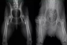
The first radiological symptoms of the disease are a slight slant (flattening) of the outer-lateral part of the affected epiphysis and rarefaction of its bone structure with an expanded radiographic joint space.
Somewhat later, the “wet snow” symptom is revealed, which consists of the appearance of heterogeneity in the bone structure of the epiphysis with areas of increased and decreased optical density and indicates the development of osteonecrosis.
This is followed by the stage of impression fracture, which has a more distinct radiographic picture and is characterized by a decrease in height and compaction of the bone structure of the epiphysis with the loss of its normal architecture - the symptom of "chalk epiphysis".
Often, the onset of the impression fracture stage is characterized by the appearance of a subchondral pathological fracture line in the affected epiphysis - the "nail" symptom, the localization and length of which can be used to predict the size and localization of a potential focus of necrosis - sequestration, and, consequently, the severity of the disease.
It is generally accepted that the first stage of the disease - the osteonecrosis stage - is reversible and with a small focus of necrosis, which is quickly revascularized, it does not progress to the impression fracture stage. The appearance of a subchondral pathological fracture line in the epiphysis indicates the onset of a long-term staged course of the pathological process, which can last for several years.
Recently, MRI has been frequently used for early diagnostics of osteochondropathy of the femoral head. This method has high sensitivity and specificity. It allows to detect and determine the exact size and localization of the necrosis focus in the femoral head several weeks earlier than it is detected on an X-ray.
Sonography also allows early suspicion of the disease, but in the diagnosis of Perthes disease it has only an auxiliary value. Sonography determines changes in the acoustic density of the proximal metaepiphysis of the femur and joint effusion. In addition, it helps to track the dynamics of restoration of the epiphysis structure.
The clinical and radiological picture of Perthes disease at subsequent stages (impression fracture, fragmentation, restoration and outcome) is typical, and diagnosis of the disease is not difficult, however, the later the diagnosis is established, the worse the prognosis regarding the restoration of normal anatomical structure and function of the hip joint.
How to examine?
Who to contact?
Treatment Legg-Calve-Perthes disease
Patients with osteochondropathy of the femoral head require complex pathogenetic treatment in conditions of complete exclusion of load on the affected leg from the moment of diagnosis. In most cases of the disease, treatment is conservative. However, in case of a large focus of necrosis involving the lateral epiphysis in children aged 6 years and older, it is advisable to perform surgical treatment against the background of conservative measures. This is due to the pronounced deformation of the femoral head and the protracted (torpid) course of the disease. Severe deformation of the femoral head, in turn, can cause the formation of extrusion subluxation in the affected joint.
Necessary conditions for complex pathogenetic treatment:
- elimination of compression of the hip joint caused by tension of its capsular-ligamentous apparatus and tension of the surrounding muscles, as well as continuing axial load on the limb;
- changing the spatial position of the pelvic and/or femoral components of the affected joint (using conservative or surgical methods) with the aim of completely immersing the femoral head into the acetabulum, creating a degree of bone coverage equal to one;
- stimulation of restorative processes (revascularization and reossification) and resorption of necrotic bone tissue in the femoral head, freed from compressive influences and immersed in the acetabulum.
Conservative treatment
Conservative treatment is carried out under bed rest conditions, with the affected lower limb placed in a position of abduction and internal rotation, facilitating full immersion of the femoral head into the acetabulum. This position is supported by a Mirzoeva splint. A plaster bandage-spacer on the knee joints according to Lange, cuff or adhesive plaster traction for the thigh and shin, as well as some other devices that also perform a disciplinary function.
The required abduction and internal rotation in the hip joint is usually 20-25°. The Mirzoeva splint and cuff traction are removed for the duration of medical and hygienic measures - usually no more than 6 hours per day. Traction is also performed around the clock in courses lasting 4-6 weeks, coinciding with physiotherapy courses, at least 3-4 courses per year.
The advantages of removable devices are the possibility of full-fledged therapeutic gymnastics and physiotherapy procedures. In addition, it becomes possible to walk on crutches on a limited basis without support on the sore leg or with a measured load that helps stimulate the reparative process at the recovery stage, and it makes it easier to care for the patient. However, in the absence of proper control over the child's stay in such devices, it is recommended to apply a plaster cast in the Lange position. The child's ability to move with crutches depends on the patient's age, development of motor coordination and discipline. The nature of the lesion is also important - unilateral or bilateral.
Often, the beginning of treatment under the conditions of a centering device is prevented by chronic sluggish synovitis of the hip joint, accompanying Perthes disease - painful limitation of abduction and (or) internal rotation of the hip, and in some cases - the formed vicious position of flexion and adduction.
In case of inflammation of the affected joint, drug treatment with NSAIDs - diclofenac and ibuprofen in age-appropriate dosages and anti-inflammatory physiotherapy are used to restore the range of motion of the hip. The duration of such treatment is usually 2 weeks. If there is no effect, tenomyotomy of the contracted subspinal and/or adductor muscles of the hip is performed before applying a plaster cast or abduction splint.
Therapeutic gymnastics is an important part of the treatment and consists of passive and active movements in the hip (flexion, abduction and internal rotation) and knee joints. It is continued even after the full range of hip movements has been achieved. During physical exercises, the child should not feel any significant pain or fatigue.
Physiotherapeutic procedures - electrical stimulation of the gluteal muscles and thigh muscles, various types of electrophoresis, exposure to the hip joint area with the Vitafon vibroacoustic device, warm (mineral) mud. Thermal procedures on the hip joint area (hot mud, paraffin and ozokerite) are completely excluded.
Physiotherapy is carried out in combination with massage of the muscles of the hip joints in courses of 8-12 procedures at least 3-4 times a year.
Electrophoresis of angioprotectors on the spine area is combined with electrophoresis of angioprotectors and microelements on the hip joint area, as well as with oral administration of osteo- and chondroprotectors. Electrophoresis of the ganglion blocker azamethonium bromide (pentamine) is prescribed for the thoracolumbar spine (Th11-12 - L1-2), aminophylline (euphylline) for the lumbosacral spine, and nicotinic acid for the hip joint area. Electrophoresis of calcium-phosphorus-sulfur, calcium-sulfur-ascorbic acid (using the tripolar method) or calcium-phosphorus is prescribed for the hip joint area.
Control radiography of the hip joints in the anteroposterior projection and Lauenstein projection is performed once every 3-4 months. The question of putting the child on his feet without supporting means is decided upon completion of the radiological stage of recovery.
In almost all cases of the disease in children under 6 years of age, the prognosis with conservative treatment is favorable - the significant potential for new bone tissue formation in the affected femoral head and the growth of its cartilaginous model ensures complete restoration of the shape and size of the femoral head (remodeling) according to the shape and size of the acetabulum. The duration of conservative treatment at this age is no more than 2-3 years.
Surgical treatment
Reconstructive surgical interventions for the treatment of children with Perthes disease:
- medializing and corrective osteotomy of the femur;
- rotational transposition of the acetabulum, which is performed both as an independent intervention and in combination with medializing osteotomy of the femur.
Among the varieties of rotational transpositions of the acetabulum, the most popular is the Salter operation.
Surgical intervention is performed with the aim of centering (complete immersion) of the femoral head in the acetabulum, reducing the compressive effect of the muscles of the hip joint and stimulating the reparative process.
The high efficiency of remodeling operations in the most severe cases of Perthes disease - subtotal and total lesion of the epiphysis has been proven by extensive clinical experience. Surgical intervention ensures a more complete restoration of the shape and size of the femoral head, as well as a significant reduction in the duration of the disease - the patient is put on his feet without supporting means on average after 12±3 months, depending on the stage of the disease.
Использованная литература


 [
[