Medical expert of the article

New publications
X-ray of fingers: indications, how to do it
Last reviewed: 04.07.2025

All iLive content is medically reviewed or fact checked to ensure as much factual accuracy as possible.
We have strict sourcing guidelines and only link to reputable media sites, academic research institutions and, whenever possible, medically peer reviewed studies. Note that the numbers in parentheses ([1], [2], etc.) are clickable links to these studies.
If you feel that any of our content is inaccurate, out-of-date, or otherwise questionable, please select it and press Ctrl + Enter.
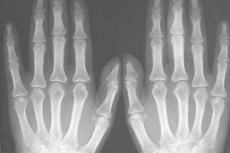
Radiography or x-ray of fingers – obtaining a fixed black and white image of their bones and soft tissues – is a method of radiation diagnostics, widely used in clinical traumatology, orthopedics and surgery.
Indications for the procedure
In most cases, a simple radiograph is the main examination in diagnosing finger injuries and pathologies of their joints, which allows you to accurately determine the location of fractures of the fingers or toes and assess their morphology (for example, transverse, oblique, spiral, with displacement, with fragments), and in the case of dislocation - to identify any displacement, compression or deviation of the joints.
In addition, indications for radiography of fingers include diagnostics of:
- joint inflammation (arthritis);
- inflammation of the periosteum - periostitis;
- inflammation of the joint capsule - bursitis of the finger;
- hallux valgus;
- deep (bony) panaritium of a finger or toe;
- local osteoporosis;
- bone and fibrous ankylosis;
- defects (abnormal growth) of bones and bone neoplasms.
In complex cases, as well as when surgical intervention is necessary, they resort to more modern and informative methods - computed tomography or magnetic resonance imaging.
Preparation
No preliminary preparation is required for X-rays of fingers and toes, but rings should be removed from fingers before the procedure.
However, if it is impossible to do this, for example, due to severe swelling of the injured finger, the X-ray is still taken: the outline of the jewelry will be visible on the X-ray, and the X-ray technician will make a corresponding mark on it. If there is a fixing plaster cast on the limb at the time of the X-ray, the image is taken through it.
Children undergo X-rays with enhanced protection, covering certain parts of the body with a lead apron.
Technique finger x-rays
Typically, an X-ray of a finger on the hand is done in the following projections:
- direct projection or anteroposterior image - a picture from the back of the hand (the hand is placed on the X-ray cassette lying on the table, palm down, fingers are held in an extended position);
- lateral projection – a picture taken from the side (the hand is placed on the edge).
The thumb is examined in a direct projection, for which the hand is turned so that the dorsal side of the finger is flat on the X-ray plate. In a lateral image, the other fingers are moved toward the elbow joint - for the most flat position of the thumb.
If it is necessary to confirm a fracture, an image is taken in an oblique projection - at an angle, which provides a clearer image of the phalanges of the fingers and increases the accuracy of the diagnosis. To create an inclination and support, the fingers in this position are placed on a stand at an angle of 45°. [ 1 ]
Standard toe x-rays are performed in frontal, lateral, and angled projections. The frontal projection requires the patient to lie on his or her back, with the knees bent and the feet flat on the table. Lateral and angled toe x-rays are performed similarly to foot x-rays, with the patient positioned in the same way.
What does a finger fracture or dislocation look like on an x-ray?
A finger fracture on an X-ray looks like an uneven, lighter strip (line or gap) against the background of the bone, often with displacement of fragments or angular position of the fragments.
A dislocation of a finger on an X-ray shows a shift (displacement) of the surfaces of the metacarpophalangeal or interphalangeal joint, that is, the separation of its head from the socket - complete or partial. In the latter case, a subluxation is diagnosed. [ 2 ]
See also - X-ray signs of damage to bones and joints