Medical expert of the article

New publications
X-rays of the sacroiliac joints
Last reviewed: 03.07.2025

All iLive content is medically reviewed or fact checked to ensure as much factual accuracy as possible.
We have strict sourcing guidelines and only link to reputable media sites, academic research institutions and, whenever possible, medically peer reviewed studies. Note that the numbers in parentheses ([1], [2], etc.) are clickable links to these studies.
If you feel that any of our content is inaccurate, out-of-date, or otherwise questionable, please select it and press Ctrl + Enter.
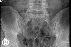
X-ray of the iliosacral (iliosacral) joints visualizes the most important anatomical structures of the musculoskeletal system: the paired sacroiliac joints, which connect the articular surfaces of the sacrum (os sacrum) and the ilium bones (os ilium) that are part of the pelvic ring.
Indications for the procedure
Visualization of these joints using X-rays is carried out:
- in case of injuries to the sacroiliac joint (one or both) and pelvic bones – cracks and/or fractures; [ 1 ]
- to determine the cause of frequent or persistent sacroiliac joint pain, felt as pain in the lower back (lumbosacral spine) or pelvic area;
- in cases of problems with movement due to a violation of their stability (these joints are classified as partially mobile joints - amphiarthrosis);
- for the purpose of diagnosing local inflammatory processes, for example, if sacroiliitis is suspected.
Preparation
Before this examination, preparation is required, which consists of temporary (for three days before the procedure) restriction of food consumption rich in fiber (cellulose), as well as increasing gas formation in the intestine. And if there is such a problem as constipation, laxatives should be used during these same three days.
In addition, the evening before the X-ray, you should not eat after 7 p.m., and in the morning you should do a cleansing enema.
Technique X-rays of the sacroiliac joints.
Conventional radiographic technique involves shielding the body parts adjacent to the X-ray exposure zone: when examining the iliosacral joints, lead plates, according to the radiation protection protocol of the International Commission on Radiological Protection (ICRP), should protect the upper abdomen.
The specific location of the structures of the sacroiliac joint - the parts of the ilium and sacrum that form it are located at an angle to the sagittal (middle) plane of the body, overlapping each other in the frontal (straight) plane - requires targeted radiography in several projections.
The patient is placed on the X-ray table on his back, but the part of the body below the waist should be at a slight angle to the horizontal surface of the table, for which purpose rollers are used. The cassette with the X-ray film is placed where the upper posterior protrusions (spines) on the crest of the iliac spines are projected, and the beam of the X-ray machine is focused from a distance of one meter on the area of the abdominal cavity - slightly to the side of its midline, at the level of the upper anterior spines os ilium. [ 2 ]
The patient's position is semi-sitting and the body is tilted forward or backward (with the cassette placed under the buttocks) may be necessary in cases of fractures of the iliosacral joints. [ 3 ]
Complications after the procedure
A short-term negative consequence of this examination may be a feeling of discomfort and increased pain in the inflamed or injured joint. To avoid this, a local anesthetic (novocaine block) may be administered before the procedure.
There have been no recorded cases of complications after this procedure, since the radiation doses are very low, and when the total dose is less than 1000 mSv (millisieverts), there are no health risks.
For comparison: when X-raying the bones of the pelvic ring (including the sacrum) in a direct projection, the radiation dose does not exceed 2.23 mSv, in a lateral projection – 1.57 mSv.
Reviews
Numerous reviews from specialists indicate that the diagnostic capabilities of X-rays of the sacroiliac joint for identifying the causes of the so-called sacroiliac pain syndrome are quite limited: according to estimates, the accuracy of this method does not exceed 40.5%, and the sensitivity does not reach 30%.
Radiography is also not suitable for the early detection of sacroiliitis and other lesions of the sacroiliac joints, so other instrumental diagnostic methods are used, in particular, osteoscintigraphy, computed tomography or magnetic resonance imaging.