Medical expert of the article

New publications
X-rays of the wrist joint for children and adults
Last reviewed: 03.07.2025

All iLive content is medically reviewed or fact checked to ensure as much factual accuracy as possible.
We have strict sourcing guidelines and only link to reputable media sites, academic research institutions and, whenever possible, medically peer reviewed studies. Note that the numbers in parentheses ([1], [2], etc.) are clickable links to these studies.
If you feel that any of our content is inaccurate, out-of-date, or otherwise questionable, please select it and press Ctrl + Enter.
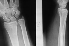
Since X-rays provide an image of the dense structure of bones, X-rays of any joints, including X-rays of the wrist, are a classic method for the initial diagnosis of joint injuries and diseases.
Indications for the procedure
An X-ray of the wrist joint (which connects the hand to the forearm and consists of eight bones) is mandatory in cases of acute or chronic wrist pain - often with joint contracture and other symptoms that may arise as a result of:
- bone cracks or fractures;
- joint dislocation;
- bone hypoplasia or the presence of additional bone structures;
- arthrosis or arthritis; [ 1 ]
- periostitis (pathological changes in the periosteum) and osteochondrosis of the wrist;
- joint deformities associated with osteoarthritis, osteophyte formation or calcifications. [ 2 ]
More detailed information in the material - Causes of pain in the wrist joint.
Examination of the wrist joint using X-rays makes it possible to visualize bone structures (ligaments and muscles are not visible on X-rays) and identify existing deviations, as well as conduct differential diagnostics, establishing the etiology of damage and confirming the diagnosis.
In addition, based on an X-ray examination of the wrist and hand, the results of treatment are assessed, in particular, bone fusion (formation of bone callus) in fractures.
X-rays of the uninjured wrist are also necessary before surgery for carpal tunnel syndrome.
Technique X-rays of the wrist joint.
For general and targeted fluoroscopy of the hand and its wrist joint, the technique is identical; for details on protecting other parts of the body, read the publication - X-ray of the hand.
However, some features of hand positioning in cases of wrist fracture should be taken into account. To obtain the most informative images, images are needed both in direct projection (from the back side - with ulnar deviation of the wrist, from the palm side - with bent phalanges of the fingers), and in lateral projections - with the wrist tilted forward and backward. And in case of a fracture affecting the scaphoid bone of the joint, also in oblique projection.
The conclusion that the X-ray of the wrist joint is normal is made when, upon studying the obtained X-ray images and comparing the image with the norm, no pathological changes in the articular bone structures are revealed, that is, their location and the spaces between them correspond to the normal anatomy of the wrist joint.
X-ray signs of wrist fracture
As already noted, X-rays for the most common fracture of the wrist joint – its scaphoid bone – are taken in different projections on the arm bent at the elbow. [ 3 ]
The diagnosis of a wrist fracture is confirmed by visual signs, in particular:
- in case of a fracture without displacement – the presence of a strip of light on the line of damage to the bone;
- destruction of the outer (cortical) layer of bone;
- displacement of bones, causing deformation of the wrist;
- the presence of bone fragments or comminuted fragments in a segmented fracture.
Each X-ray image – in accordance with the protocols available to radiologists – is accompanied by a detailed description of all the characteristics of the identified pathological changes in bone structures (with measurements of bone displacement parameters and the localization of their fragments in millimeters and degrees). [ 4 ]
Reviews
As radiologists themselves note, fluoroscopy of the wrist joint cannot always reveal its pathological changes. The same fracture affecting the scaphoid bone is difficult to visualize using X-rays, so additional diagnostic examinations are often used - MRI and osteoscintigraphy.