Medical expert of the article

New publications
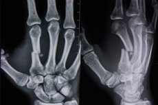
X-ray of the hand
Last reviewed: 06.07.2025

All iLive content is medically reviewed or fact checked to ensure as much factual accuracy as possible.
We have strict sourcing guidelines and only link to reputable media sites, academic research institutions and, whenever possible, medically peer reviewed studies. Note that the numbers in parentheses ([1], [2], etc.) are clickable links to these studies.
If you feel that any of our content is inaccurate, out-of-date, or otherwise questionable, please select it and press Ctrl + Enter.
X-ray examination is considered a fairly common diagnostic method, which is used to determine the state of the musculoskeletal system, detect disorders, injuries and diseases. Among other types of X-ray diagnostics, many patients are prescribed X-ray of the hand - this is a painless and relatively quick procedure that has a minimal radiation load on the human body, and therefore is safe. Specialists can prescribe X-rays of the hand 3-4 times a year, if necessary: this will not cause any harm to health.
Indications for the procedure
According to statistics, X-rays of the hands are most often performed in cases of injuries, painful processes, and other conditions that negatively affect the functionality of the upper limb, as well as its configuration (which is usually noticeable from the outside).
The following are considered basic indications for performing an X-ray:
- pain of varying intensity in the area of the hand, both after physical activity and at rest;
- joint deformities;
- violation of bone integrity, bruises;
- inflammation in the hand area, tumor processes (both benign and malignant);
- joint defects – for example, of congenital etiology (in Turner syndrome). [ 1 ]
Using an X-ray of the hand, it is often possible to make the following diagnoses:
- cystic formation (benign cystic tumor localized in the center or in the subchondral part of the bone epiphysis);
- synovitis (accumulation of effusion in the joint cavity);
- tenosynovitis, tendinitis (inflammatory process in the tendon and synovial tendon sheath);
- calcification (deposition of calcium salts, one of the symptoms of arthritis);
- osteophyte (a spiky bony growth at the border of the articular surface); [ 2 ]
- osteoporosis (a disease associated with the loss of calcium from the bones).
According to a 2013 study, DXR (digital radiographic) analysis of wrist and hand X-rays can predict the risk of hip fracture in women and men.[ 3 ]
X-ray of the hand for bone age
When speaking about bone age, doctors mean a conditional age period corresponding to the level of development of the skeletal system. It is usually determined using X-rays, after which special calculation indicator schemes are used: the person's body weight and height, chest circumference and stage of puberty are taken into account. [ 4 ]
There are several methods for assessing the bone-age index. These methods take into account the period of appearance of the epiphyseal sections of tubular bones, the stages of their development, the stage of joining the epiphyses and metaphyses with the formation of synostoses. The above processes are especially well visible in the bones of the hands of the upper limbs, since they have a considerable number of epiphyseal sections and ossification nuclei.
The level of skeletal maturity can essentially be determined based on two characteristics: the rate of growth in the areas undergoing ossification and the rate of calcium accumulation in these areas. From infancy to adulthood, these two characteristics follow a specific pattern and time schedule. [ 5 ] The timing of epiphyseal ossification and bone fusion does not occur uniformly throughout the body. In some bones, ossification begins immediately after birth, while in others it occurs between 14 and 17 years. [ 6 ]
Determination of bone age is usually performed in cases of physical development disorders in children, slow growth, pathologies of the pituitary gland, hypothalamus and thyroid gland.
Preparation
X-rays of the hands do not require any special preparation, regardless of which hand will be examined – the right or the left.
Immediately before the procedure, the patient must remove metal jewelry: rings, bracelets, wristwatches must be removed. The patient must sit on a chair in the X-ray room, placing the hand on a special support: the position of the limb will be indicated by the specialist who will conduct the diagnosis.
If necessary, the patient may be given special protection in the form of a lead apron or vest.
Technique X-rays of the hand
X-ray of the hand is performed as follows. The patient sits on a chair located near the table or couch of the X-ray machine. Usually, the specialist asks to bend the arm at the elbow joint, placing the hand on the table or a special stand. The angle of the hand will be indicated by the radiologist. The following types of images are most often used:
For direct projection, the hand is placed horizontally on the surface of the support. In this case, the X-rays will pass through the hand perpendicularly, which will allow the entire bone system of the wrist to be examined, except for the pisiform bone. The metacarpal bones, carpometacarpal joints, finger phalanges, and intercarpal joints are well visualized.
For the lateral projection, the palm is placed with the lateral edge on the surface, and the thumb is moved forward. This position facilitates good examination of the bone contours, phalanges, and metacarpal bones. The lateral projection is often used to assess the degree of wrist injury, since displacements of bone segments are well visualized.
For the oblique dorsal projection, the hand is placed on the dorsal surface at an angle of 45°. This angle will help to examine the condition of the first and fifth metacarpal bones, as well as the triquetral, hamate and pisiform bones.
For the oblique palmar projection, the hand is positioned with the palmar surface at an angle of 45°. This allows visualization of the trapezoid and scaphoid bones.
Sometimes the position of the brush is adjusted individually, depending on the existing problem.
X-rays of the right hand are usually taken in two projections for optimal examination of the problem. The hand is placed as flat as possible on the table surface, with the fingers pressed together. X-rays of the left hand are taken in the same way, and only in some cases is an atypical positioning of the limb used, which is determined individually.
X-rays of the fingers help to give an objective assessment of the structure and condition of bones, soft and cartilaginous tissues. The doctor individually determines the need to obtain an image of several or all fingers of the hand - usually in two projections. The patient's task is to hold the fingers still during the procedure. If such immobility cannot be ensured, then additional fixation is used.
Often, along with the examination of the injured or pathologically altered limb, an X-ray of the healthy hand is performed for comparison.
X-ray of the hand of a child
X-rays of the hand are allowed for children of any age, if there are compelling indications. For the youngest patients, it is convenient to use a special "X-ray cradle" - a device in which it is possible to fix the child to obtain a clear image. If there is no such cradle, then the baby should be held by the mother or someone close to her, because it is impossible to obtain a high-quality image during movement.
If possible, it is advisable to have a digital X-ray of the child’s hand: it is safer and more comfortable for the child.
Diagnostics in childhood may be prescribed:
- in case of traumatic injury to the upper limb and wrist area;
- in case of insufficient development of limbs, as well as for establishing bone age;
- for local pain in the hand;
- in tumor processes, developmental anomalies of bones and wrist joint.
- to assess the bone age of a child. [ 7 ]
Fracture of the wrist on x-ray
The hand has a complex structure, as it consists of a large number of small and tiny bones. Therefore, fractures here are varied and often multiple. On an X-ray, fractures can be represented by the following varieties:
- open and closed fractures;
- complete or partial fractures (cracks);
- single or multiple bone injuries;
- diaphyseal, periarticular or extraarticular fractures;
- primary or secondary fractures;
- with or without displacement of fragments.
Identifying a wrist fracture on an x-ray is relatively easy if the limb is positioned correctly before the image is taken.
X-ray of wrist dislocation
A dislocation is a displacement of the articular surfaces of the bones of the hand relative to each other, which can be caused by trauma or other pathology. X-ray examination of a dislocation plays one of the main roles: it can be used to determine the degree of injury and exclude other bone damage. The image can easily identify the type of dislocation, which can be true, perilunate, periscaphoid-lunate, transscaphoid-perilunate, peritriquetral-lunate, transscaphoid-translunate, depending on the position of the hand during the injury and the direction of the force impact.
X-ray of the hand is prescribed if a dislocation is suspected, if the victim fell, leaning on the hand, or received a direct blow to the wrist area. As a rule, the X-ray is performed in two, and if there is any doubt, in three projections.
X-ray of the hands in rheumatoid arthritis
Hand radiography is the most valuable imaging modality in rheumatology. Joint disease can be identified by individual features such as joint space narrowing, erosion, subluxation, and deformity. In diseases such as rheumatoid arthritis, the presence of erosions on hand radiographs provides valuable information about disease progression and response to therapy. [ 8 ]
An X-ray examination is especially necessary for a patient with rheumatoid arthritis – first of all, to assess the extent of the pathological process and determine its stage.
The basic radiographic signs of the disease are edematous soft articular tissues against the background of a slight narrowing of the interarticular space and visible osteoporosis. In case of long-standing pathology, bone erosions will be determined - small defects of the edges of the bone articular ends. The epiphyses of the bones of the finger phalanges are usually distinguished by circular enlightenment.
If an X-ray of the hands with scaling was performed, the signs will be more extensive: a rupture of the occlusal plates is detected, and almost at the very beginning of the development of the pathology. Over time, the X-ray picture worsens: the joint narrows, erosive foci form, osteoporosis becomes more distinct. With the destruction of the final joint elements, subluxations may develop.
Contraindications to the procedure
X-ray diagnostics has long been the only method that allows you to look "inside" the human body. However, this procedure has quite a few contraindications, so over time, specialists have developed new methods for conducting research - in particular, magnetic resonance imaging and ultrasound.
Contraindications to x-rays of the hands are:
- pregnancy period (this contraindication is relative, since with proper protection the study can still be carried out);
The dose of ionizing radiation required to cause specific effects on the fetus depends on the stage of pregnancy. [ 9 ] The U.S. National Council on Radiation Protection states that the risk of miscarriage or major congenital malformations in fetuses exposed to doses of 5 rad or less is negligible compared with the spontaneous risk among unexposed women. The spontaneous risk includes a 15% chance of spontaneous abortion, a 3% risk of major malformations, and a 4% risk of fetal growth retardation. [ 10 ], [ 11 ]
The Centers for Disease Control and Prevention's Committee on Radiation Safety recommends that unborn children of laboratory workers should not be exposed to more than 0.5 rads of cumulative radiation during pregnancy.[ 12 ]
- lactation period;
- psychopathologies (manic psychosis, schizophrenia, etc.);
- decompensated, severe conditions of the patient.
It is better not to take X-rays too often, so as not to exceed the permissible radiation dosage. It is also not recommended to resort to X-ray diagnostics without special indications. [ 13 ]
Normal performance
The radiologist performs a description or decoding of the image immediately after receiving the image. This process involves assessing the relative position of the bones, the state of their connections and integrity, structural features and the degree of density.
The norm is when the bones have a homogeneous structure. X-ray images should not have dark spots on a white background, and gaps are required between bone elements.
In case of traumatic injury to one hand, an X-ray of the other hand may be required to conduct a comparative analysis and more easily identify deviations.
Complications after the procedure
If it is possible to choose an X-ray machine for examining the hands, then preference should be given to a modern digital device: its radiation dose is lower than that of earlier generation analogs.
For each diagnostic procedure involving X-rays, the doctor registers the patient in the dose load log and also makes a note in the individual outpatient card. Just a few years ago, the radiation dose was calculated using a special table that indicated average values. Today, any modern X-ray machine or tomograph has a built-in sensor that immediately after the procedure shows the dose level that the patient received. This dose - for example, when performing an X-ray of the hand - cannot be the same for everyone. It depends on the area of the study, the hardness of the rays used, the distance from the emitter, etc.
Usually, it takes a fraction of a second to take a picture of the hand. During this time, no negative consequences for the body can occur. However, in some cases, additional protection in the form of lead aprons, plates and collars is still required. For example, such protection is necessary if the study is performed on a pregnant woman or a child.
To avoid negative consequences, it is also important to remember that the level of radiation can accumulate, so it is not recommended to take one shot after another in a short period of time: the body must have time to recover.
It is not recommended to have X-rays of the hands during pregnancy, especially in the first trimester. However, in some cases – for example, if the integrity of the bones is compromised – it is impossible to do without an X-ray. To avoid possible complications for the growing fetus, protection is used: special screens in the form of aprons, which cover the woman’s chest and abdominal area from radiation.
According to sanitary standards, the maximum permissible dose of radiation for a fetus is determined by an indicator not exceeding 1 mSv. At the same time, the average dose during an X-ray of the hand is usually less than 0.1 mSv, so it cannot cause any harm.
Experts advise not to panic in vain, but to have an X-ray of the hand if there are really indications for this procedure. The radiation load will be minimal, and the diagnostic information that the doctor will receive will be comprehensive: the doctor will be able to establish the correct diagnosis and prescribe the correct treatment. [ 14 ]
Care after the procedure
Special patient care after the procedure is usually not required. Immediately after the examination, the doctor will decipher the results, send the results to the attending specialist, or prescribe treatment independently. The patient, depending on the circumstances, will be sent home or to the hospital for further treatment.
Some doctors recommend drinking plenty of fluids and eating dairy products: milk, kefir, natural yogurt, to reduce radiation exposure on the day of the procedure. In addition, it is advisable to take a shower immediately upon arrival home. It is better to diversify your diet with fruits and greens, natural freshly squeezed juices. And we must not forget that an X-ray of the hand is a safe diagnostic, so there is no need to worry about possible long-term negative consequences.