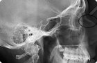
There are several methods of X-ray: without the use of a contrast agent (overview image) and with its use, allowing to monitor its progress within the kidneys, ureter, bladder.
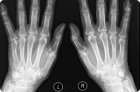
X-ray of hands does not require advance preparation. The only requirement is the absence of metal objects on them: rings, bracelets. In the presence of gypsum at the time of radiography, he is removed.
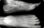
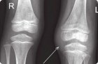
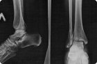
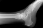
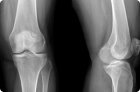
The most accessible, informative and painless methods of visualization of bone structures is radiography. The picture also shows damage to the joints, cartilage traumatic and inflammatory genesis, and birth defects.
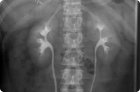
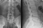
The most accessible diagnosis, which allows visualizing the internal structure and assess the state of the skeletal bones of the spine, is radiography.
Radiography refers to the methods of radiation diagnosis and is a non-invasive examination of the internal structure of a certain part of the body by radiating it with X-rays and obtaining a projection of the image on a special film.
During the procedure, several photographs are taken simultaneously. All is determined by how much movement is carried out in the joint being examined. The method used is called double contrasting.
The most widely used non-invasive diagnostic method for detecting congenital and acquired pathological changes in bone and joint tissues is the visualization of their anatomy with the help of X-rays.
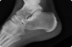
Pain in the knee, impaired mobility of the joint in this area and traumatic injuries are quite frequent reasons for contacting a doctor. Determine by eye, with which unpleasant symptoms are associated, it is not easy even for an experienced doctor.