Medical expert of the article

New publications
X-rays of the hand, fingers, forearm and upper arm: how are they done?
Last reviewed: 03.07.2025

All iLive content is medically reviewed or fact checked to ensure as much factual accuracy as possible.
We have strict sourcing guidelines and only link to reputable media sites, academic research institutions and, whenever possible, medically peer reviewed studies. Note that the numbers in parentheses ([1], [2], etc.) are clickable links to these studies.
If you feel that any of our content is inaccurate, out-of-date, or otherwise questionable, please select it and press Ctrl + Enter.
It is difficult to imagine medicine today without the discovery of the German physics professor Wilhelm Roentgen, who, while studying electric rays, discovered that they penetrate dense material and project its image onto a screen. He first saw an X-ray of his hand, placing it in the path of the rays. Replacing the screen with a photographic plate, he gave the world his discovery in the form in which it exists to this day. Without it, ultrasound, MRI, and CT would be impossible. For what medical indications is an X-ray of the hand prescribed now?
Indications for the procedure
The need for an X-ray of the hand arises when the patient complains of pain in the limb, a fall or other injury has occurred, or changes in the appearance of the hands have been noted. In this case, the doctor assumes the following diseases:
- rheumatoid arthritis is an inflammation of the joints of the extremities, most often the hands. It starts with the smallest ones, gradually affecting the cartilage and leading to deformation of the articular bones. X-ray of the hand gives a picture of the degree of damage and violation of the integrity of the bone;
- polyneuropathy - damage to the structure of the nerve fibers of the peripheral nerves of the motor system, disruption of the process of muscle contraction. Expressed in numbness of the hands, tingling, sometimes pain;
- arm fracture - a traumatic injury that leads to a violation of the integrity of the arm bone in any of its segments. Most often, the lower third of the radius, the phalanges of the fingers, and the metacarpal bones are subject to fractures;
- shoulder fracture - injuries to the shoulder, especially its neck, are not spared. They are most often characteristic of elderly people;
- dislocation of the arm - to recognize it and differentiate it from fractures if the usual clinical examination methods are not enough. X-rays reveal the non-adjacent joint surfaces, possible complications, obstacles to reduction and its outcome.
X-rays are also necessary for shoulder osteosynthesis - the introduction of metal structures to restore its anatomical integrity (pins or plates are used). X-rays are used to monitor the healing of the bone wound.
Preparation
X-ray of hands does not require preliminary preparation. The only requirement is the absence of metal objects on them: rings, bracelets. If there is a plaster cast at the time of the X-ray, it is removed.
Pregnant women should inform their doctor about this, as he may choose a safer method of examination for the fetus, such as MRI, which provides a clearer picture and does not use radiation.
Technique X-rays of the hand
X-ray of each area of the hand requires its own technique; in each case, for greater information content, one or another angle is required, its various projections: direct, lateral, oblique palmar and dorsal.
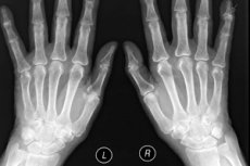
X-ray of the hand
To do this, a person sits on a chair near the device. The arm is bent at the elbow, and the hand is on the table; it must be completely immobilized during the shooting. The rays pass perpendicular to the hand to obtain a direct projection, so the wrist bones are visible.
Lateral projection is needed to detect bone displacements of the wrist, phalanges, metacarpal bones. The image is obtained using a cassette on which the palm is placed sideways, with the thumb slightly abducted.
The oblique palmar is necessary to determine the condition of the trapezoid and scaphoid, trapezium bones. The image is taken on a cassette, with the palm raised by 45 0 in relation to it.
Oblique dorsal - allows viewing the first and fifth metacarpal, triquetral, pisiform, hamate bones. The algorithm of this projection is similar to the previous one, only the palm is placed with the back side.
 [ 1 ]
[ 1 ]
X-ray of a finger on a hand
In case of a finger fracture, an X-ray helps to establish the nature of the damage, its location, where the bone fragments are displaced, if any. The film should show the 2 closest joints, so the image is taken in several projections and repeats the first 2 points of the X-ray of the hand.
After performing surgical or conservative treatment for complex injuries, a control X-ray is taken, then repeated after 10-12 days, when the swelling has subsided, and also after the plaster is removed.
X-ray of the bones of the forearm
Carrying out an X-ray of the forearm requires, first of all, its complete immobility, the slightest tremor can distort the picture. For a complete image, two angles are necessary: direct and lateral projection, the field of view also includes the scapula with the collarbone.
The procedure is performed in a sitting position, sideways to the device. The hand, forearm and shoulder are exposed. The arm, bent at the elbow, is placed on the table with the palm facing up to obtain a direct projection. A lateral projection is obtained by placing the palm with the edge to the surface.
X-ray of the shoulder
For an X-ray examination of the shoulder, you need to undress to the waist. It is carried out in two projections lying on the table; if for some reason it is impossible to take this position, the image is taken sitting or standing.
As a rule, adults are given an image of only the injured shoulder, and children are given an image of both the sick and healthy shoulders to compare the development of bone tissue.
[ 2 ]
X-ray of a child's hand
Due to radiation, X-rays of the hands of children and pregnant women are performed only according to strict indications and very carefully, and their frequent repetition is avoided.
Often, an endocrinologist prescribes an X-ray examination for children, this is due to delayed or accelerated growth. X-rays reveal the "bone" age and bone growth reserve.
To do this, they take a picture of the hand and the lower third of the wrists, since it is easiest to take pictures of the upper limbs. By comparing the data with the standards, they identify the pathology that needs to be dealt with before puberty.
X-ray of hand at home
Modern medicine is able to provide X-rays at home for elderly people and patients with functional disorders. For this purpose, there is a portable device, with the help of which a picture of various organs, including the shoulder, forearm, and hand, is taken at home.
The images are developed on site, printed, described and given to the patient, and based on the results, specialists carry out treatment.
Complications after the procedure
Each fluoroscopy procedure carries a small dose of radiation, which, if certain rules are followed, will not cause any negative consequences. To do this, you need to use shielding of body parts that are not subject to examination, use time protection, i.e. do not resort to the procedure often, but only when indicated.