Medical expert of the article

New publications
Ankle x-rays
Last reviewed: 06.07.2025

All iLive content is medically reviewed or fact checked to ensure as much factual accuracy as possible.
We have strict sourcing guidelines and only link to reputable media sites, academic research institutions and, whenever possible, medically peer reviewed studies. Note that the numbers in parentheses ([1], [2], etc.) are clickable links to these studies.
If you feel that any of our content is inaccurate, out-of-date, or otherwise questionable, please select it and press Ctrl + Enter.
Nowadays, traumatology increasingly encounters various injuries. One of the weakest organs that is most often subject to injury is the foot. It is quite easy to damage it. This does not require heavy falls, blows, or displacements. A light push is enough, and the foot will be damaged. Also, sometimes it is only necessary to twist your leg a little - and the foot will also be injured. This is especially true for women, since they often wear heels. Professional athletes also often encounter such injuries, for example, when performing physical exercises without a preliminary warm-up, or in a mode of increased load, overfatigue. In this regard, one of the most popular diagnostic methods at present is an ankle X-ray.
Indications for the procedure
An ankle X-ray may be prescribed primarily for diagnostic purposes, in order to establish the type and nature of the injury to which the ankle has been subjected. It is used to diagnose fractures, joint displacements, dislocations, cracks, sprains, ruptures and tears of ligaments. It is also used to examine the foot to establish the causes of swelling, hematomas, injuries, internal defects and bleeding.
The need for this type of X-ray also arises in connection with changes in normal mobility, shape, structure, appearance of the foot, as well as with intense pain in the ankle area. With the help of X-rays, it is possible to confirm diagnoses such as arthritis, arthrosis, diagnose various types of inflammatory processes, edema. The procedure also allows you to identify neoplasms, deformations.
Preparation
The procedure does not require lengthy preparatory activities (in terms of adherence to a specific diet, rest, work, physical activity). Preparation can be reduced exclusively to psychological and moral preparation for the upcoming examination.
The doctor must explain to the patient who, how, and for what purpose will conduct the examination, as well as talk about the expected expectations from the procedure. The patient must have a rough idea of the procedure, understand its essence and significance. He must also have an idea of the purpose for which it is carried out, what risks arise in connection with the procedure.
When conducting the examination, the victim must take the necessary position on the couch. The laboratory assistant or doctor conducting the examination must tell or show the patient the position he must take. The legs must be bent at the knees, and the feet must be placed on a flat surface. If it is necessary to identify an ankle injury, the X-ray is taken in the lateral projection. For this, the patient must be seated. The injured limb must be placed on a support.
In order to determine the degree of transverse or longitudinal flatfoot, it is necessary to provide increased load on the ankle joint. When examining the arch of the foot, the patient should stand on one leg and tuck the other.
It is also necessary to collect a preliminary anamnesis during the preparation process. For example, if an X-ray procedure was already performed 6 months ago, then the X-ray cannot be repeated, since this is associated with a high level of radiation exposure to the body. It is also important to inform the doctor about pregnancy and breastfeeding, since this is a contraindication for the procedure. The exception is cases of severe injuries. In this case, a special lead apron is required, which can provide protection from radiation.
Ankle joint placement
To conduct the examination, it is necessary to correctly position the ankle joint. First, a visual assessment of the injury is carried out, and then an X-ray examination is performed. The procedure as a whole takes no more than 10 minutes.
The most commonly used projection is the direct posterior projection. The advantage of this projection is that it eliminates rotation of the foot. To perform the examination in this position, the patient must lie down, with the legs extended along the surface of the table, horizontally. The sagittal plane of the foot must form an angle of 90 degrees with the surface of the table.
The examination can also be performed in a direct posterior projection with foot rotation. To do this, the leg must be positioned in the same way as it is positioned during the examination in a direct posterior projection (lying down, legs along the table). The difference is that the foot must be turned inward, creating an angle of 15-20 degrees.
When examining the ankle joint in the lateral projection, the patient is positioned in a lying position on his side. The limb that is not being examined should be pressed to the stomach, and the foot of the other surface should be in contact with the lateral surface. In this case, the heel should be pressed tightly to the cassette, the foot should be turned inward by approximately 15-20 degrees.
The examination can be carried out with or without load on the foot.
Technique ankle X-rays
The essence of the study is that X-rays are passed through the tissues that need to be examined. They pass through soft tissues and are retained by hard tissues. The image shows differences between healthy and damaged tissues, as well as between soft and hard tissues. Modern equipment makes it possible to obtain high-quality images with well-visualized complexes.
The examination is carried out in various projections. Most often, direct and lateral projections are used. The patient is placed in the required position, then the examination itself is carried out, passing X-rays through the tissues being examined. The ankle must be fixed in a position that maximally visualizes the area that needs to be examined.
X-ray of the outer ankle
The procedure should be performed in a lying position. In this case, it is mandatory to keep the body part being examined immobile. The patient is placed in one of three possible positions and the leg is fixed in such a way as to maximally visualize the required area.
Contraindications to the procedure
Contraindications include pregnancy and lactation. Age under 15 is also a contraindication. The study should also not be performed frequently, the frequency should not exceed 3-4 times a year.
Ankle X-ray During Pregnancy
X-ray examination is contraindicated during pregnancy, especially in the first trimester. The lactation period is also a contraindication. Since at this time the body receives radiation exposure (radiation), which is dangerous for both the mother and the fetus. Radiation can cause various genetic and somatic mutations in the fetus, developmental defects, serious damage to the nervous, hormonal, and immune systems. Sometimes the fetus can even die. Radiation is especially dangerous in the first trimester, since during this period the fetus has minimal protection and is most vulnerable. X-rays are contraindicated during lactation due to the fact that radiation can accumulate in breast milk.
However, despite the contraindications, the procedure can be performed in any of the above cases if there is an urgent need. In this case, to minimize radiation, it is necessary to use a special protective apron or protective pads. For example, pregnant women use pads on the abdomen and pelvic area.
Normal performance
In a healthy person, the soft tissues and bone tissues are visualized separately in the image. Homogeneity is observed: the rupture lines are not reflected, the tissue looks uniform and homogeneous.
Normally, there should be an angle between the foot and the shin. Normally, it should be 130 degrees. It may be lower, but exceeding these indicators indicates pathology. The arch of the foot is normally 35 mm or more.
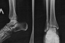
Ankle fracture on x-ray
If a fracture is suspected, the doctor will order an X-ray examination. In this case, it is most often necessary to compare the healthy leg and the damaged one. The fracture is clearly visible on the image as a pronounced fracture line located on the bone tissue.
Complications after the procedure
The procedure does not have serious consequences. No damage occurs, the load on the body does not increase. The exception is cases of frequent procedures. X-rays cannot be taken more than once every 6 months. This is due to the fact that each procedure is associated with a certain level of radiation. Negative consequences may also be observed when the procedure is performed on pregnant women. Radiation can cause mutations, gene defects.
There are no known cases of complications after X-rays. Theoretically, it is believed that X-ray radiation can cause an allergic reaction in people with increased allergy. However, there is no data to support this theoretical position.
Care after the procedure
The procedure does not require any special care. After a person has had an ankle X-ray, he or she can return to their daily activities immediately after the procedure.
 [ 24 ]