Medical expert of the article

New publications
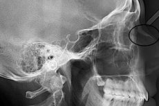
X-ray of the sinuses and nasal bones in a child and adult
Last reviewed: 03.07.2025

All iLive content is medically reviewed or fact checked to ensure as much factual accuracy as possible.
We have strict sourcing guidelines and only link to reputable media sites, academic research institutions and, whenever possible, medically peer reviewed studies. Note that the numbers in parentheses ([1], [2], etc.) are clickable links to these studies.
If you feel that any of our content is inaccurate, out-of-date, or otherwise questionable, please select it and press Ctrl + Enter.
Radiography is a method of radiation diagnostics and is a non-invasive study of the internal structure of a certain part of the body by shining X-rays through it and obtaining a projection of the image on a special film. This is one of the main diagnostic examinations that has entered medical practice since the end of the last century and is still relevant today due to its availability and high information content. X-rays of the sinuses and nasal bones are prescribed after an injury, if there is a suspicion of an acute disease of this localization, a neoplasm, or to monitor the results of treatment.
Ionizing radiation during examination can indeed have a harmful effect on the body, and this is known to all. However, is an X-ray of the sinuses harmful? And to what extent?
Passing through the tissues of a living organism, X-rays ionize neutral atoms and molecules, turning them into charged particles. However, the danger is primarily in long-term exposure to radiation, as well as intensive exposure. Diagnostic equipment uses short-term low-intensity irradiation. It is considered practically safe even when repeated many times.
Moreover, we do not undergo nasal X-rays as often as, for example, fluorography, so if necessary and in the absence of contraindications, there will be no harm from a one-time procedure, even if after some time you are prescribed another control study.
An X-ray of the nose is necessary for an otolaryngologist to assess the condition of the bone structure of the nose and surrounding tissues, the degree of their damage, in order to establish the correct diagnosis and not make a mistake in choosing the method and tactics of treatment.
How often can you get an x-ray of your sinuses and nasal bones?
The maximum permissible total annual dose of radiation received from all sources is considered to be 150 mSv (milliSieverts). Such a dose can be received by a person if regular radiation diagnostics are necessary for vital indications (about 100 examinations per year).
If there is no such need, then over the course of a year the average citizen will accumulate a dose in the range of 5-15 mSv.
A single X-ray of the sinuses on the most modern digital equipment will result in 0.12 mSv of radiation, while on the most "shabby" one it will be 1.18 mSv. So even several examinations, if necessary, will not result in a lethal dose for the patient.
It is considered undesirable to undergo more than two examinations per year, because X-rays of other parts of the body may also be needed. However, the frequency of X-rays will be determined by your attending physician, in this matter you will have to trust him, because the assessment of the effectiveness of the treatment is of great importance to confirm its correctness. For example, patients with a fracture of the nasal bones with displacement have to regularly monitor how the recovery process is going, and several unscheduled diagnostic procedures will cause much less harm than incorrect blind treatment.
Indications for the procedure
An X-ray examination of the paranasal sinuses is prescribed if the following symptoms are present, which allow one to suspect the presence of an inflammatory process:
- nasal congestion that makes breathing difficult, prolonged runny nose;
- periodic nosebleeds;
- a feeling of distension, heaviness in the paranasal sinuses, photophobia, lacrimation;
- a sharp increase in temperature or persistent subfebrile temperature without apparent reason;
- swelling and redness of the skin in the nose area;
- pain in the forehead, which intensifies when trying to tilt the head toward the chest.
X-ray of the nose in case of sinusitis and other inflammatory processes in the sinuses of the nose allows us to determine the pathological accumulation of a liquid substance in them and differentiate the localization of inflammation, for example, ethmoiditis (inflammation localized in the ethmoid labyrinth) from frontal sinusitis (damage to the frontal sinus) or sinusitis.
In addition, radiography of the sinuses and nasal bones can diagnose:
- the presence of a foreign object in the nose;
- tumors, cysts, polyps, papillomas;
- deviated nasal septum;
- osteomyelitis;
- osteoporosis.
An X-ray of the nose is mandatory if there is a suspicion of fractures or cracks in the nasal bones due to bruises and blows to the facial part of the head. It is necessary to visualize the type of damage to the nasal bones, the presence of displacements, and to determine the urgency of providing assistance. For example, an X-ray will show the presence of such a dangerous complication of a fracture as air entering the frontal part of the skull. In this case, every hour counts. Therefore, if you receive an injury, you should not delay going to the X-ray room.
An X-ray can detect deviations from the norm at any stage of their development: hematomas, fractures and other destructive changes in the nasal bones, damage to the nervous tissue and blood vessels. Even if you did not have an X-ray of the nose immediately after the injury, it is never too late to do so, especially if you are concerned about discomfort or feel a disturbance in breathing.
Nasal X-rays are prescribed to patients before planned surgical interventions in this part of the skull in order to visualize the anatomical features of this area that may become an obstacle to performing a standard operation.
Preparation
No special preparation is required before the X-ray examination. It is necessary to warn the doctor about the presence of metal non-removable objects in the examination area, for example, dental crowns, and remove metal jewelry (chains, remove the ring from the nose).
In the X-ray room, patients are given special vests with lead plates sewn into them for the duration of the procedure, so as not to irradiate other parts of the body unnecessarily.
To obtain a clear image, the patient must take a certain position and remain still for several seconds.
 [ 4 ]
[ 4 ]
Technique X-rays of the sinuses and nasal bones.
The sinuses or paranasal sinuses are located in the facial and partially cerebral bones of the skull. The epithelial surface of the sinuses is a continuation of the mucous membrane of the nasal passages. X-rays of the paranasal sinuses are taken in the nasomental, chin and axial projections, each of which is used to visualize a specific anatomical structure. Sometimes additional positions are used to examine defects in more detail. The final choice of projection remains with the radiologist, who can make adjustments to the otolaryngologist's appointment.
When examining the sinuses, the patient assumes a vertical (standing or sitting) or horizontal (lying) position, depending on the capabilities of the available equipment.
The maxillary or maxillary sinuses are located, as their name suggests, in the body of the upper jaw. X-rays of the maxillary sinuses are taken in most cases in the chin projection. In the image from this position, they are shown most openly. Most often, during this procedure, the patient sits or stands near a vertical X-ray stand, sometimes the patient is laid on a table.
In the nasomental projection, the pyramids of the temporal bones prevent a clear view of the maxillary sinuses along their entire length, blocking the lower third of the view, and sometimes completely closing it. To neutralize this visualization defect, when performing an X-ray of the maxillary sinus in this projection, the patient is asked to open his mouth during the shooting, while the temporal bones are lowered down, opening the view. To detect fluid in the maxillary sinus, the image is taken in a vertical position. If such measures are not enough, then a maxillary sinusography is performed - an X-ray with the introduction of a contrast agent into the maxillary sinuses. This method allows you to detect formations inside the sinuses - polyps and cysts. Maxillary sinusography of the left and right sinuses is done alternately, and not simultaneously.
An X-ray of the frontal sinuses is prescribed if frontal sinusitis is suspected. It is performed in a direct projection centered on the frontal bone, under which these sinuses are located. The patient stands with his chin resting on a special support. The radiologist or lab technician helps him to take the correct position. Sometimes an X-ray in this projection is performed in a lying position.
The images of the posterior nasal cavities are taken in the axial projection, which clearly shows the sphenoid and ethmoid sinuses, and the rocky part of the temporal bone, the openings of the base of the skull and damage to these bones, if any, are clearly visible in the image in this projection. If defects are detected in the image in the axial projection, additional targeted, clearer radiographs are taken if necessary. Also, the lateral position of the patient can be used to visualize the paranasal sinuses.
X-ray of the nasal septum allows to detect its curvature, congenital or acquired, in time. Such pathology causes a violation of the nasal breathing function and increases the likelihood of developing sinusitis. The curvature of the nasal septum is clearly visible in the images in the nasofrontal projection.
X-rays of the nasal bones are usually performed in direct (nasochin or nasofrontal) and lateral (right or left) projections. The diagnostic procedure is performed as soon as possible after receiving a blow to the face.
A direct projection image shows only fractures with displacement. To determine the sides of the injury, the patient is laid on his side on each side; sometimes it is necessary to take an image in the nasomental projection, where the structure of the nasal bones and maxillary processes is clearly visible.
In case of impression fractures (when only transverse displacements occur), the images are taken in axial projection. This displacement is also detected on a targeted X-ray of the frontal sinus, where the nasal passages are clearly visible.
Special categories of patients
X-rays of the sinuses during pregnancy are performed only in cases of extreme necessity. The pregnant woman must cover her abdomen with a protective lead vest.
X-rays of the paranasal sinuses in children are also performed only in cases where the benefits of the procedure outweigh its harmful effects, since X-rays have a negative effect on the development of bone tissue. Indications for X-rays of the paranasal sinuses in children include facial injuries, suspected foreign body in the nose, curvature of the nasal septum, suspected inflammation of the paranasal sinuses, congenital anomalies of the nasal structure, adenoids. The child should have the following symptoms:
- noisy breathing, snoring, sleep disorders;
- nasal congestion and voice changes;
- elevated temperature;
- headaches;
- developmental disorders of the facial bones of the skull.
An alternative diagnostic method for a child is magnetic resonance imaging, which is permitted from birth and does not involve radiation exposure. However, its availability is limited.
In a child, the interosseous sutures of the facial bones are clearly visible and have a cartilaginous structure. In cases of minor bruises, they diverge to the sides, but their integrity is not violated. In childhood, the following traumatic disorders of the nasal bone structure are common: the introduction of bones between the frontal processes and flattening of the nasal vault overhang. Their visual symptom is the recession of the nasal bridge, an increase in the distance between its bones - the nose becomes flattened, the edges of its bones may protrude. In such cases, radiography is not informative; rhinoscopy is used, which allows identifying hematomas and tissue ruptures.
Contraindications to the procedure
An absolute contraindication to the procedure is a severe mental disorder, in which the patient cannot fulfill the conditions necessary for the procedure: take the necessary position, hold his breath, and so on.
If there are metal fixed prostheses in the area of transillumination, it is recommended to replace radiography with another visualization study.
X-rays are contraindicated for pregnant women due to their teratogenic effects, and for preschool and primary school children due to their negative impact on skeletal growth and development.
For people with weakened immunity, routine diagnostics may be postponed until a more favorable period.
Emergency radiography for vital indications is performed on almost all categories of the population, observing safety precautions.
Normal performance
An X-ray can provide almost complete information about the condition of the paranasal sinuses and nasal bones, identify the presence of an inflammatory process, neoplasms, damage to bone and cartilage tissue, and also establish that everything is in order with regard to the patient’s respiratory system.
An X-ray of the sinuses of a healthy person is characterized by clear lines and contours of the bones, smooth contours of the paranasal sinuses, and the absence of thickening of the mucous membrane enveloping the bone walls. Absolute symmetry of the nasal sinuses is not required.
The paranasal sinuses should contain only air, their color on the X-ray is light gray, comparable to the color inside the eye sockets (it is a standard for comparison). The patient has a smooth nasal septum, intact bones and clearly visible contours of the ethmoid cells.
What does an x-ray of the sinuses show, decoding
Nasal X-rays can detect various signs of diseases. Their description with a presumptive diagnosis usually takes the radiologist about ten minutes. Several images taken dynamically can also be used to track positive changes in the treatment process or their absence. In case of inflammation of the nasal sinuses, several images are usually prescribed: diagnostic and for monitoring the treatment. Decoding an X-ray of the nasal sinuses includes not only a description of their condition, but also deviations from the norm of other anatomical structures that are visible in the image. Sometimes asymptomatic pathologies are accidentally discovered in this way, for example, neoplasms or forgotten incorrectly healed fractures that have led to bone defects.
Darkening of the sinus compared to the standard indicates the presence of inflammation (sinusitis). X-ray clearly shows its localization: in the frontal part (frontal sinusitis); maxillary sinuses (sinusitis); sphenoid (sphenoiditis), ethmoid cells (ethmoiditis). Often, several paranasal sinuses are involved in the inflammatory process: bilateral - hemisinusitis, affecting all sinuses - pansinusitis.
In addition, the X-ray image can accurately determine the type of inflammatory process: simple or catarrhal, serous, purulent, exudative. These processes differ only in the type of substance accumulated in the sinuses, which is determined by puncturing. The accumulation of fluid looks like a darker than air area with an upper horizontal level. Sometimes the border of the liquid substance has the form of a parabolic curve with the apex below. This shape indicates a violation of the communication of the sinus with the nasal cavity.
Also, literally by two X-rays, it is possible to distinguish an acute process from a chronic one. To do this, when performing a repeat X-ray, the patient's head is moved to any side. In case of acute inflammation, the fluid boundary will also shift, in case of chronic inflammation - it will not.
Parietal hyperplastic sinusitis, as well as polypous sinusitis, are clearly visualized. The first form is characterized by darkening along the contour of the walls of the nasal sinuses. This occurs because a hyperplastic process occurs in the mucous membrane covering the bone walls, due to which it thickens. The contour of the sinuses in this case is directed inside the sinus and has an uneven or wavy edge. In advanced cases, the sinus is completely darkened and turns into an airless space.
A nasal polyp or multiple growths of it visually look like a protrusion of the wall on a stalk, facing into the sinus.
Tumors appear as darkened areas. A cyst is visualized as a faint or more pronounced round shadow, bordered by a smooth, clear line.
Neoplasms are usually discovered unexpectedly. They do not have any pronounced clinical signs except for frequent inflammations in the nasal sinuses and some difficulty breathing. When detected, surgical treatment is prescribed.
[ 10 ], [ 11 ], [ 12 ], [ 13 ]
Broken nose
X-rays of a broken nose can determine fracture lines, the presence of displacement of fragments and splinters, as well as their presence in soft tissues and sinuses, and help assess the degree of damage to the perinasal tissues. Minimal damage is an isolated fracture of the nasal bone without displacement.
X-ray diagnostics for a nasal fracture is a highly informative method that allows detecting fractures and cracks at early stages, when the inflammatory process has not yet affected soft tissues to a significant extent. This method is also important for monitoring the process of bone fusion and callus formation.
Radiography can help determine the type of fracture: straight, oblique or transverse; multi-fragmented or bird's beak; no displacement; differentiate the fracture from a deviated nasal septum.
Fractures are also classified according to the mechanism of injury, which is important for forensic examination.
A picture in the nasomental projection allows us to detect such a complication as hemorrhage into the sinuses.
Sometimes, an X-ray of the skull and sinuses reveals an “air bubble sign” – a complication in the form of air entering the frontal part of the skull. In the image, it is visible under the cranial vault and frontal bones.
The anatomical features of the organ are of great importance. If the nose has a thin and short structure, then the line of enlightenment (fracture) may be outside the resolution and not be determined.
Large and long nasal bones are damaged more often, and the resulting defects are very clearly visible on the image.
Mild nasal bone injuries are characterized by a crack and hemorrhage in the quadrangular cartilage; a fragile lower edge. The bones are deformed in this case, and a curvature of the pyriform aperture is observed.
If the blow to the nose came from the side, the X-ray shows the displacement of both bones. The picture resembles a fracture, but the line of enlightenment and displacement of bone fragments are not visible.
Fractures of the nasal bones from a side impact appear as widening of the anterior bones, since the crack occurs at the junction of the nasal bones with the eye sockets.
A blow from top to bottom is characterized by: an impression and/or vertical fracture of both nasal bones; deformation of the frontal processes. In case of damage to the cartilaginous tissue, the line of enlightenment (fracture) is often not visible, since this type of tissue is characterized by elasticity. However, with this direction of the blow, a crack in the quadrangular cartilage occurs, and also - a displacement of the septum is possible. There are many nuances that can be visualized using targeted radiographs.
Complications after the procedure
Medical examinations using X-rays use low-intensity radiation for several seconds. X-rays of the sinuses and nasal bones are among the shortest and safest in terms of the dose of radiation received among X-ray examinations of different parts of the body. Even with multiple repetitions of this diagnostic procedure, no immediate consequences can arise after the procedure. And long-term consequences, for example, the risk of developing cancer in the future in people who have undergone this examination and those who have never been subjected to it, are almost the same.
The carrier of radiation in diagnostic equipment are electromagnetic waves that disappear immediately after the procedure. They are not capable of accumulating in the body, like radioactive chemicals, so no measures to remove radiation after an X-ray examination are required.
However, you should only undergo X-ray examinations as prescribed by your doctor and monitor the amount of radiation you have received throughout your life.
So, we have found out that complications do not arise after the X-ray procedure. However, refusal of diagnostics can lead to serious consequences, the mildest of which is a deviated nasal septum. Without visualization of lesions of any genesis, nasal diseases are complicated by the development of respiratory failure, suppuration of the muscles and tissues of the face, as well as infection of the brain. It is possible to "overlook" hematomas, neoplasms, hyperplasia. Inadequate treatment leads to chronic inflammation, constant swelling of the soft tissues of the face.
Analogs to X-ray
An alternative radiation diagnostic method is computed tomography. Unlike an X-ray, the doctor receives a clearer three-dimensional image that can be copied to a laser disk or flash drive, or sent by e-mail. However, computed tomography gives the most significant radiation exposure. The radiation dose during computed tomography of the skull and paranasal sinuses is 0.6 mSv. Compared to a modern X-ray machine, this is true: when taking one X-ray, you will receive 0.12 mSv. Even if you take it in two projections. On antediluvian equipment, the dose received will already be 1.18 mSv, with two projections - twice as much. So the radiation exposure from CT is not always greater than that of an X-ray. The price of the issue is the cost of the procedure.
Examination of internal organs using ultrasound waves (echosinusoscopy) is considered the safest, it is recommended even for pregnant women – an unborn child is subjected to ultrasound scanning. However, some organs remain partially inaccessible to ultrasound scanning. Among them are bone tissue and paranasal sinuses, since they normally contain air. Ultrasound diagnostics are available for the frontal and maxillary sinuses of the nose, it can detect neoplasms and the presence of fluid or foreign bodies. Ultrasound can diagnose a deviated nasal septum. However, this method, in addition to its main advantage – safety, also has a number of disadvantages. Ultrasound data often lead to hyperdiagnosis (can indicate a pathology that does not exist), so many doctors still require a clarification of the diagnosis using an X-ray. X-rays are considered more informative. Ultrasound is often prescribed as an additional research method for examining the anatomical structures of the nose, not excluding X-rays.
Magnetic resonance imaging is quite informative and is considered safe. It can also be used to diagnose injuries and diseases of the nose. However, in addition to the high cost of the study, radiation methods (X-ray and computed tomography) are considered more informative when examining the bone structures of the facial skeleton. MRI better visualizes soft tissues, vessels and nerves, as well as neoplasms in them.
Various methods can be used to examine nasal structures, but X-rays are the most versatile and informative, and, importantly, accessible.
Reviews of the procedure are the most favorable, it is short-term, does not cause any unpleasant sensations and the patient's condition before and after the procedure does not change. Due to the cheapness of radiography, the presence of radiological laboratories in almost all outpatient departments, as well as high information content, it is very common. The only advice that "seasoned" patients give is to, if possible, do X-rays in rooms equipped with the most modern equipment. It has many advantages - from the comfort of the patient and higher-quality images to the lowest radiation doses.