Medical expert of the article

New publications
Joint arthrography
Last reviewed: 04.07.2025

All iLive content is medically reviewed or fact checked to ensure as much factual accuracy as possible.
We have strict sourcing guidelines and only link to reputable media sites, academic research institutions and, whenever possible, medically peer reviewed studies. Note that the numbers in parentheses ([1], [2], etc.) are clickable links to these studies.
If you feel that any of our content is inaccurate, out-of-date, or otherwise questionable, please select it and press Ctrl + Enter.
Arthrography is an X-ray examination method that examines a joint. The examination is performed immediately after a contrast agent is injected into the joint. Sometimes air is also injected. Often, both the contrast agent and air are injected at the same time. Together, they create the ability to visualize the contours of the joint structures formed by soft tissues. It also becomes possible to examine the joint surface.
During the procedure, several images are taken simultaneously. Everything is determined by the range of motion in the joint being examined. The method used is called double contrast.
Indications for the procedure
Arthrography is recommended for persistent pain in the joint area. However, the procedure is also indicated in case of joint dysfunction of unclear etiology. This may be bacterial, viral, fungal joint damage. The procedure is also performed for allergic joint damage, in cases of frequent crepitus, and joint injuries. Indications include various autoimmune joint damage, inflammatory and degenerative processes.
Preparation
Preparation does not require any specific techniques. The essence of preparation is that the essence of the study being conducted is explained to the person, as well as its principles, purpose, objectives, and expected results (what the study will presumably show). It is important to inform the person who, where, and how the study will be conducted.
Preparation does not imply the need for any additional restrictions in the diet, work and rest regime. No specific diet is required either. The patient must understand that the essence of the study is that, using X-ray methods, they look at the features of the movement of the contrast agent through the joints. They control that the substance completely fills the joint cavity and also begins to distribute over the joint tissue. After the substance has completely distributed, X-rays are taken.
The patient should be warned that local anesthesia is used. It is advisable to conduct a study in advance for the presence of an allergic reaction. An immediate reaction is considered especially dangerous, especially anaphylactic shock. It is also worth collecting anamnesis to predict possible reactions of the body to the administration of anesthesia, to the contrast agent.
The patient must be warned that the examination may cause some discomfort, despite the fact that local anesthesia is used. He must understand that the examination may be accompanied by a feeling of pain, discomfort, tingling or a feeling of distension in the joint area.
The patient should also not move during the examination. The number of involuntary movements should also be kept to a minimum. The person should not move unless the person conducting the examination gives the appropriate command.
It is also necessary to understand that the patient must strictly follow the doctor's instructions, especially if they concern changing the position of the body, movements. The instructions should be followed as quickly and clearly as possible, which will allow the examination to be performed as clearly and as quickly as possible.
You should not drink large amounts of liquid for several days before the examination, and your food should be moderate. You should stop taking medications containing iodine and its individual components. You should also warn the doctor in advance about any current illnesses, since it is necessary to find out exactly whether the patient has health problems that could complicate the procedure in some way.
 [ 6 ]
[ 6 ]
Technique arthrography
There are several ways to perform an X-ray examination of joints using the arthrography method.
The first, main method of arthrographic examination is considered to be the method in which a contrast agent is injected into the joint cavity. Most often, a contrast agent containing iodine or other iodine-containing compounds is used. This method is called "positive contrast". Air is also used as a contrast agent. In this case, we are talking about negative contrast.
The second method is also considered to be a method based on a mixture of air and a radiopaque substance, which acts simultaneously as a positive and negative contrast.
The use of each method has its own distinctive features and its own scope of application. For example, in case of damage to the joint capsule and ligaments, it is advisable to resort to the use of the positive contrast method. In cases where the meniscus is torn, or a cartilaginous defect is noted, it is better to use the second method. When examining children, during routine and preventive examination of adults, for a gentle examination of adults and the elderly, this method is also used. It is necessary to take into account that if it is necessary to perform a puncture, sterile conditions are required.
If fluid accumulates in the joint capsule, it is necessary to pump out this fluid before introducing the contrast. The amount and size of the intervention depends on the size of the composition. It is necessary to take into account that the contrast agent is absorbed very quickly. It is advisable to take X-rays immediately after the contrast agent is introduced, otherwise the image contours will be unclear and blurred.
The technique and specifics of the procedure may vary. Thus, depending on the specific type of research that needs to be conducted, different research techniques are used.
For example, the shoulder method of examination has its own specifics, which is used in the diagnosis of a rupture of the cuff muscles. The rotator cuff of the shoulder is a group of muscles that is located at the highest point of the shoulder. It is often advisable to examine this area in the process of diagnosing a shoulder dislocation. The results of the examination make it possible to obtain important diagnostic information about the condition of the joint capsule, as well as about the features of pathological changes in the area of the biceps tendons.
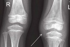
When examining the knee joint, meniscus injury is considered as the main indication for the examination. This method is very reliable. Thus, its reliability in detecting injuries is 90%. Also, with the help of arthrography, it is possible to diagnose Baker's cysts, which are cyst-like growths that form from synovial fluid and are often localized in the area of the synovial sac. These growths can form as a result of bulging of the synovial membrane against the background of weakening of the knee joint.
Meniscus injuries often occur against the background of chronic polyarthritis or meniscus damage, weakness of the knee joints. Arthrography makes it possible to diagnose changes and defects in the knee joint area. However, in case of cartilage and ligament injuries, this procedure is uninformative.
The elbow diagnostic method is a diagnostic method that is used when there is a suspicion of a ligament rupture, as well as determining the location of joint mice.
The radiocarpal method is a method for diagnosing injuries and ligament ruptures, and is also used for symptoms of inflammatory processes in the area of the articular ligament.
In the anterior metatarsal form, arthrography is used to diagnose various injuries of the joint capsule. It is used for rupture of external and internal ligaments. Arthrography of the shoulder and knee joint is the method that is most often used in diagnostics.
Examination of other joints does not have significant diagnostic value.
TMJ arthrography
This involves examining the temporomandibular joint. This form of arthrography involves examining the joints, which involves creating artificial contrast in the cavity being examined and then conducting further examination using radiography.
It is advisable to conduct the examination in the morning, on an empty stomach. First, the skin around the site of the proposed puncture is treated. Sanitary and hygienic procedures and antiseptic measures are mandatory. Preliminary treatment involves washing, removing hair, and also direct treatment of the puncture site with an antiseptic. Regular alcohol is used as the main antiseptic. Then local anesthesia is administered in the form of a 1% solution of novocaine. Penicillin or other antibiotics are administered to prevent joint infection.
There are three options for conducting arthrography studies.
In the first case, nitrous oxide or oxygen is blown into the joint cavity. This method is called pneumoarthrography.
In the second case, a heavy high-atom contrast agent is injected into the joint – this is the high-atom arthrography method.
In the third case, the method of double contrasting of the joint is used, which involves the introduction of both gas and a heavy contrast agent into the joint cavity.
After the procedure, you are asked to make certain movements in the joint, after which X-rays are taken in various projections. If necessary, the computed tomography method is also used.
[ 7 ], [ 8 ], [ 9 ], [ 10 ], [ 11 ]
Arthrography of the shoulder joint
When examining the shoulder joint, the joint is treated for disinfection. Antiseptic solutions are used for this purpose. Various local anesthetics are injected subcutaneously. Additionally, to prevent infection and to reduce the pain threshold, an anesthetic is injected directly into the head of the humerus. For this purpose, the needle is inserted directly into the joint capsule and advanced until it feels abutted against the articular cartilage.
After removing the mendren, a syringe containing a contrast agent is attached to the needle. Under the control of fluoroscopic techniques, 1 ml of contrast agent is injected into the joint cavity, and the needle is slowly pulled towards you. If the needle is positioned correctly (and this will be visible on fluoroscopy), then the remainder of the contrast agent can be injected. After this, the needle is slowly removed from the joint, and a sterile swab is used to remove the remainder. After the needle has been removed, a series of images are quickly taken. This allows for high image quality and good clarity.
CT arthrography
Using computer tomography methods (or, as it is also called, CT arthrography), it is possible to obtain a detailed image of the joints subject to contrast. In this case, the contrast is introduced in the usual traditional way. With this method, it is possible to examine small parts and cavities. It is usually used to examine small parts that cannot be diagnosed using X-rays. In this case, the need for a puncture of the contrast agent disappears. No special preparation is required.
[ 15 ], [ 16 ], [ 17 ], [ 18 ], [ 19 ], [ 20 ]
MR arthrography
Magnetic resonance imaging, which is based on the introduction of a contrast agent. This is the most informative method of the entire line of studies. This technique allows you to visualize those parts of the joint that cannot be seen using other research methods. For example, using this method, you can diagnose capsules or cavities, various intra-articular and extra-articular surfaces. It is used to make a diagnosis, to diagnose articular cartilage, meniscus, various inflammatory and infectious diseases.
Arthroscopy
It is used mainly to diagnose the condition of large joints. Arthroscopy is used to examine the knee, hip, and shoulder joints. It makes it possible to identify various pathological diseases, including those of inflammatory genesis. Arthroscopy can be used to track parameters over time. It makes it possible to diagnose pathological changes in the biceps and rotator cuff muscles. This diagnostic method is uninformative when examining small joints.
[ 21 ], [ 22 ], [ 23 ], [ 24 ], [ 25 ], [ 26 ], [ 27 ]
Fistulography
This method allows you to evaluate the shape, size, and current condition of the articular surfaces, soft tissues, and bones. The indicators can be evaluated to obtain information about processes occurring dynamically or statically. There are practically no contraindications.
Contraindications to the procedure
Arthrography is not performed during pregnancy and breastfeeding. It is also advisable to postpone the use of this method of examination in the acute stage of arthritis, it is worth waiting at least for the condition in which arthritis from the acute form passes into the usual form.
Contraindications include infectious joint disease, blood clotting disorders, skin diseases, external skin and mucous membranes.
The method may also be contraindicated in case of a tendency to allergic reactions. In particular, a strict contraindication is an allergic reaction to iodine and iodine-containing compounds.
Complications after the procedure
Usually the procedure is quick, side effects and negative consequences are rare. Pain may occur during the injection (administration of the drug), and may also persist for some time after the procedure (at least the first 1-2 hours). In exceptional cases, allergic reactions occur, especially if the person has a tendency to hypersensitivity and anaphylaxis.
The main complications are considered to be the development of inflammatory processes that arise as a consequence of the intervention. This may be an individual reaction of the body, or a consequence of incorrect or unclear administration of the drug. Against the background of increased sensitization of the body, an allergic reaction may develop, the severity of which can vary widely, ranging from an allergic rash, burning, irritation, and ending with anaphylactic shock.
If sanitary and hygienic requirements and aseptic rules are not observed, an infectious process, purulent-septic, inflammatory conditions may develop.
Crepitation processes, which are accompanied by a feeling of crunching, clicking when moving the joint, can also be considered as complications. A feeling of burning, distension, swelling in the joint area can also be observed for several days.
Care after the procedure
After the procedure, it is necessary to immobilize the joint that was examined. The immobilization period is 12 hours. In order to ensure the immobility of the joint, elastic bandages and bandages are applied. A special knee pad is used to immobilize the knee joint. Movements after 12 hours should be gradual and easy. To reduce swelling, ice is applied to the affected area.
If pain occurs, anti-inflammatory or painkillers should be used. If the temperature rises or a large amount of fluid is released from the joint, you should immediately consult a doctor. Also, if swelling, redness, hyperemia in the injection area develops, an immediate consultation with a doctor is also required. Physical activity should be limited for some time. Otherwise, arthrography does not require changing the usual regime.
[ 35 ]