Medical expert of the article

New publications
X-ray of the ankle joint.
Last reviewed: 06.07.2025

All iLive content is medically reviewed or fact checked to ensure as much factual accuracy as possible.
We have strict sourcing guidelines and only link to reputable media sites, academic research institutions and, whenever possible, medically peer reviewed studies. Note that the numbers in parentheses ([1], [2], etc.) are clickable links to these studies.
If you feel that any of our content is inaccurate, out-of-date, or otherwise questionable, please select it and press Ctrl + Enter.
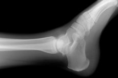
The most widely used non-invasive diagnostic method for detecting congenital and acquired pathological changes in bone and joint tissue is visualization of their anatomy using X-rays. Defects that have appeared in the skeletal structure of the foot and/or ankle can be detected by X-rays of the ankle joint, since degraded and healthy tissues absorb X-rays passing through them differently, which is reflected in the projection image of this area of the body.
Indications for the procedure
It is prescribed as part of a diagnostic examination for patients with ankle joint injuries with suspected fractures and dislocations, as well as with complaints of pain and other discomfort in this localization, which may indicate inflammatory, dystrophic and oncological pathologies.
In patients with established lesions of the joint and/or bone tissue of the ankle, radiography is performed to monitor the effectiveness of treatment.
Technique an ankle x-ray.
A little anatomy: the joint connecting the bones of the leg and foot has a rather complex structure - a system of cartilage and muscles connects three bones: the large and small bones of the leg and the calcaneus (talus) bone of the foot.
Clinical signs of ankle injuries are very similar to symptoms that occur with destructive changes in the talocalcaneal and talonavicular joints, as well as the calcaneal and calcaneal bones. Therefore, radiographs are taken in two or three projections so that these anatomical structures can be clearly seen.
The direct dorsal projection provides a good view of the calcaneal bone node and part of the tibia; the dorsal projection, with the foot turned inward, allows one to examine the tibiofibular syndesmosis (joint); the lateral projection shows the dorsal sides of the tibia, large and small.
To perform a lateral projection examination, the patient is placed on the table in a lying position on the side of the affected limb, with the limb slightly bent at the hip and knee joints. The healthy limb is pulled up to the chest as much as possible so as not to interfere with the view.
To perform radiography in a direct dorsal projection, the patient is placed on his back, bending the uninjured leg at the knee joint and pulling it towards the body. The foot of the injured leg is placed with the heel above the cassette at a right angle to the table, the outlet of the X-ray machine is directed at the ankle joint.
To control the condition of the tibiofibular joint, in the same position, the patient's foot is turned inward, the angle of rotation is approximately 30 degrees. To prevent the foot from falling down, a pad is placed under it.
Normal performance
This diagnostic method helps to identify various injuries to the joint and bone tissue of the ankle:
- injuries – closed and open fractures of bones in a given location, including cracks, complete and incomplete displacements of the bone in the joint (dislocations, subluxations);
- inflammatory processes – arthritis, osteomyelitis, synovitis, bursitis;
- degenerative changes, deformations of bone and joint tissue caused by metabolic disorders – gout, arthrosis, arthropathies;
- other congenital and acquired constitutional disorders of the articular elements.
Description of ankle x-ray
The radiologist describes the visible structural changes in the structure of the connection of the shin and foot bones, making a diagnostic conclusion. The norm of the ankle joint on the X-ray is used as a standard.
The correct proportions of the structural elements of the ankle are characterized by a uniform height of the joint space - a straight line that can be drawn through the center of the separated rounding of the tibia, as a rule, should intersect the center of the node of the calcaneus (between its elevations). Subluxation of the ankle on an X-ray usually looks like a wedge-shaped joint space. However, such an anatomical feature in rare cases is also a variant of the norm, then a similar structure of this element should be on both limbs.
The criteria for the correct positioning of the patient's leg in the direct dorsal projection are the distant parts of the tibia, the calcaneus and the X-ray joint space, the appearance of which resembles the letter "G".
In the direct dorsal projection, the calcaneus is not fully displayed. Its node is clearly visible, which should look like an irregular quadrangle with clearly visible upper and lateral sides. The upper side of the calcaneus is horizontal, slightly sagging in the middle, the medial and lateral elevations are visible, as well as the groove separating them. The plate that closes the surfaces of the joints of this connection should be clear and thin.
In this projection, the lateral process is clearly visible. The outline of the plate should uniformly pass into its contour, covered with articular cartilaginous tissue, increasing the area of the malleolar surface of the block. Its structure is spongy. All this leads to the fact that fractures of the posterior (lateral) process are intra-articular.
For a more thorough examination of the lateral part of the joint space of the ankle, a picture with the foot turned inward is examined. In it, the gap is visible along its entire length as a curved ribbon-like clearing, the shape of which resembles the letter "P".
In this same image, the tibiofibular syndesmosis can be seen more clearly; its width should normally be between four and five millimeters. The maximum permissible fluctuations in this indicator are between two and nine millimeters. The width of the soft tissues distributed over the lateral and medial surfaces should be uniform, and their volume should be small.
The dorsal part of the distal rounded end (epiphysis) of the tibia, which in surgery is often called the third (posterior) malleolus, is one of the most likely locations for a fracture, often combined with disruption of the integrity of the medial and/or lateral malleoli.
Five to six millimeters up above the top of the contour line of the medial malleolus, a horizontal line is visible against the background of the spongy formation - the outline of the notch of its dorsal section. The medial section of the distal meta- and diaphysis of the fibula is superimposed in this view on the lateral section of the distant meta- and epiphysis of the tibia. This is an area of increased load intensity, where fractures are quite common - violations of the integrity of the bone, which are easy to see in the image even for a non-specialist. Fresh injuries in the form of cracks and bone depressions are usually poorly visualized, they are better visualized several days after the injury.
A specific sign of dislocations is the displacement of bones, and an increase in the distance between the surfaces of the bones is for stretching and injury to the ligaments.
Osteoporosis, which develops due to calcium deficiency, is noticeable by increasing rarefaction (transparency) of the bone in the center and compaction of the bone borders.
Osteomyelitis of the ankle joint can be detected on an X-ray about a week after the onset of the disease. At the initial stages, the partitions between the muscles and fascia, clearly visible in the image of a healthy person, are no longer visually delineated. The border separating the muscular structure and subcutaneous tissue is also not visible, the saturation and volume of soft tissues increases. The key signs of the disease are osteonecrosis - the death of bone cell tissue, sequesters - rejection of necrotic areas.
Arthrosis of the ankle joint on X-ray looks like a modification of the thickness of the cartilaginous layer and the gap between the bone structures, as well as changes in the configuration of the endplates. The joint space is unevenly narrowed and deformed. Bony tissue growths along the edge of the joints are noticeable - osteophytes, compaction of bone tissue at the border with cartilage. Calcification of the ligaments is also clearly visible on X-rays.
Arthritis on an X-ray is characterized by widening of the joint space – a consequence of inflammatory effusion into the joint cavity.
Tumors of bone, joint and soft tissues are visualized as formations without a clear outline, extending beyond the normal structure. Destructive changes surrounding the neoplasm are characteristic.
Complications after the procedure
The procedure is non-invasive and absolutely non-traumatic, and has no consequences if certain rules are followed, in particular, not to have X-rays more than once every six months. The permissible radiation load on the body should not exceed 5 mSv. Sv is a sievert, the amount of energy absorbed by the body during irradiation. It is different for different types of X-rays. More modern equipment causes less damage to the patient's body.
The main complication after the procedure is exceeding the permissible radiation threshold.
Permanent contraindications to the examination are severe mental illnesses that become an obstacle to compliance with safety rules and the presence of metal prostheses in the area being examined.
Temporary conditions include pregnancy (X-rays are taken on expectant mothers only in cases of extreme necessity, with the abdomen covered with a lead apron) and the patient’s serious condition, which requires resuscitation measures.
For additional diagnostics, the patient may be prescribed other types of diagnostics (ultrasound, MRI, CT), which allow for further clarification of the diagnosis.
Care after the procedure
No special care is required after the procedure. Reviews of X-rays are the most favorable. If all the rules are followed, the patient is given an accurate diagnosis and treatment is prescribed quickly and inexpensively.
 [ 13 ]