Medical expert of the article
New publications
Ankle fracture with dislocation
Last reviewed: 29.06.2025

All iLive content is medically reviewed or fact checked to ensure as much factual accuracy as possible.
We have strict sourcing guidelines and only link to reputable media sites, academic research institutions and, whenever possible, medically peer reviewed studies. Note that the numbers in parentheses ([1], [2], etc.) are clickable links to these studies.
If you feel that any of our content is inaccurate, out-of-date, or otherwise questionable, please select it and press Ctrl + Enter.

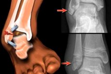
A displaced ankle fracture is defined when there is displacement of the broken bone fragments. [1]
Epidemiology
Ankle fractures are common and account for up to 10% of all bone injuries, and their incidence has been increasing in recent decades. According to foreign experts, the annual incidence of ankle fractures is approximately 190 fractures per 100,000. People, and the majority of those affected are elderly women and young men (physically active and athletes). [2] According to a nationwide population study in Sweden, closed bi- or tri- ankle fractures had an annual incidence rate of 33 per 100,000 person-years and 20 to 40 per 100,000 person-years in Denmark. [3] Interestingly, the peak incidence of trimalleolar fractures is between 60 and 69 years of age, becoming the second most common type of ankle fracture in this age group.
Supination-rotation (up to 60%) and supination-adduction (more than 15%) injuries come first, followed by injuries with excessive inward turning of the foot and simultaneous retraction or external rotation of the foot.
In this case, almost 25% of cases are fractures of both ankles (external and internal) and 5-10% are triple fractures. [4]
Causes of the displaced ankle fracture
The articular surfaces of the distal epiphyses (lower thickened parts) of the tibia and fibula (as well as the cartilage-covered convex surfaces of the body of the talus) form the ankle joint. The distal epiphysis of the tibia forms the medial (inner) ankle, and the lower part of the fibula forms the lateral (outer) ankle. Also, the posterior part of the distal end of the tibia is considered the posterior ankle.
The main causes of displaced ankle fractures are traumas of various origins (during running, jumping, falling, strong impact). There are such types as supination fractures - with excessive deviation of the foot to the outside; pronation fractures - with inward turning of the foot, exceeding the natural amplitude of movement; rotational (rotational), as well as flexion fractures - with excessive adduction and/or abduction of the foot during its forced flexion.
Most often fractures of the medial ankle, accompanied by displacement of a fragment of its part, are the result of eversion or external rotation. And a fracture of the lateral ankle with displacement can be a fracture of the fibula just above the ankle joint. This is the most common type of ankle fracture that can occur if the foot is tucked or twisted.
There can be a bimalleolar or double displaced ankle fracture - a fracture of both the lateral ankle and the medial ankle. And a displaced fracture of both ankles is considered by orthopedists to be the most severe case. And triple ankle (trimalleolar) or triple ankle fracture with dislocation involves not only the inner and outer ankle, but also the lower part of the posterior ankle of the tibia. [5]
Risk factors
Risk factors for ankle fractures include:
- Decreased bone mineral density in osteopenia, osteoporosis or hyperthyroidism;
- Increased physical stress on the ankle joints;
- Excessive body weight;
- Menopause (for women);
- Ankle joint diseases, in particular osteoarthritis, deforming osteoarthritis or tenovaginitis ankle joint;
- Weakening of the ligaments connecting the lower tibia and fibula (distal intertibial syndesmosis) associated with frequent foot tipping and ankle injuries;
- Chronic ankle instability, which develops with dysfunction of the posterior tibial tendon (and leads to acquired flat feet in adults), in the presence of diabetic peripheral neuropathy - with muscle weakness in the ankle joint and foot deformity (leading to frequent loss of balance);
- Foot malposition and foot deformities in systemic diseases.
Pathogenesis
Regardless of the localization of the fracture, the pathogenesis of the violation of bone integrity is due to the deforming effect on them of the surface energy of impact (or other mechanical action), the strength of which is higher than the biomechanical strength of bone tissue. More details on the mechanism of fracture occurrence in the publication - fractures: general information
Symptoms of the displaced ankle fracture
The clinical symptoms of ankle fracture are the same as symptoms of ankle fracture. The first signs are similar - in the form of acute pain, spilled hematoma, deformity of the ankle joint and change in the position of the foot, sharp limitation of movement of the foot with complete inability to lean on the injured leg.
Massive edema also develops very quickly after a displaced ankle fracture that involves the soft tissues of the entire foot and part of the lower leg. [6]
If the violation of the integrity of bone structures is not accompanied by soft tissue rupture, a closed fracture of the ankle with displacement of the fragments is diagnosed.
When displaced fragments break through soft tissue and skin and exit into the cavity of the resulting wound, open fracture of the ankle with displacement of the fragments is defined. In such a fracture, internal hemorrhage and bleeding of varying intensity are observed.
And violation of the integrity of the bone with more than three fragments without soft tissue rupture is a closed splinter fracture of the ankle with displacement, and with soft tissue rupture is a splinter open fracture.
Forms
A trimalleolar ankle fracture usually involves the distal part of the fibula (lateral ankle), medial ankle and posterior ankle. The first ankle fracture classification system, developed by Percival Pott, distinguished between single-, double-, and triple-ankle ankle fractures. Although reproducible, the classification system did not distinguish between stable and unstable fractures. [7], [8] Laughe-Hansen developed a classification system for ankle fractures based on the mechanism of injury. [9] It describes the position of the foot at the time of injury and the direction of the deforming force. [10] Depending on the severity of the ankle injury, different stages (I-IV) are distinguished. By providing additional information about the stability of the injury, the Laughe-Hansen classification has become a widely used classification system for ankle injuries. According to the Laughe-Hansen classification, a trimalleolar ankle fracture can be classified as SE IV or PE IV. But the Laughe-Hansen classification system has been questioned because of poor reproducibility and low inter- and intra-experimental reliability. [11]
One of the most commonly used classifications of ankle fractures is the Weber classification, which differentiates peroneal fractures related to the tibial-malleolar syndesmosis. 40 Although the Weber classification system has high inter- and intraobserver reliability, it is inadequate for multiple ankle fractures. [12]
Biomechanical and clinical studies have led to the development of classification systems for the medial and posterior ankle. Medial ankle fractures can be classified according to Herscovici et al, who distinguish four types (A-D) of fractures based on anteroposterior radiographs. [13] This is the current standard system for the medial ankle, but it is inadequate for multiple ankle fractures. [14] The indications for surgical treatment of medial ankle fractures rather depend on the degree of displacement and whether it is part of an unstable ankle fracture.
The posterior ankle can be classified according to Haraguchi, Bartonicek, or Mason. The former developed a computed tomography (CT)-based classification system for posterior ankle fractures based on CT transverse slices. [15] Mason et al modified Haraguchi's classification by specifying the severity and pathomechanism of the fracture. [16] Bartoníček et al. Proposed a more specific CT-based classification system that also takes into account the stability of the tibial-tibial joint and the integrity of the peroneal notch. [17] These posterior ankle classification systems can determine further operative or conservative treatment, but cannot fully characterize the type of triceps fracture.
The AO/OTA classification distinguishes between type A (infrasyndesmotic), B (transsyndesmotic), and C (suprasyndesmotic) peroneal fractures. [18] In addition, AO/OTA type B2.3 or B3.3 fractures are transsyndesmotic fractures of the fibula with fracture of the posterolateral margin and medial ankle. The same is true for AO/OTA type C1.3 and C2.3 fractures involving all three ankles. Additional refinements may be added to clarify the stability of the syndesmosis or associated lesions (e.g., Le For-Wagstaffe tuberosity). There is no description of the configuration of medial and posterior ankle fractures in the AO/OTA classification. This is noteworthy because posterior fragment size and displacement are factors to consider when choosing treatment. [19]
Ideally, a classification system should have high reliability between and within researchers, be widely recognized, relevant for prediction, and applicable in research and clinic. The most comprehensive classification system is the AO/OTA classification. It is widely recognized, easy to use in clinical practice, and provides information on the type of triceps fracture with emphasis on the fibula. However, an important factor, the configuration of the posterior ankle fragment, is not represented in the AO/OTA classification.
Complications and consequences
Possible complications and consequences of this type of fracture such as:
- Infection of the wound (in the case of an open fracture);
- Ankle contracture;
- Deformity of the ankle joint due to inaccurate repositioning of fragments with the development of posttraumatic arthrosis;
- Impaired reparative bone tissue regeneration leading to the formation of the so-called false joint;
- Post-traumatic habitual foot sprains;
- Improper fusion of the fracture (e.g., tilting the talus outward), making walking difficult;
- Development of impeachment syndrome of the ankle with disruption of its normal mechanics.
Diagnostics of the displaced ankle fracture
The diagnosis of ankle fracture accompanied by dislocation is determined by clinical examination.
Its main component is instrumental diagnostics, including x-ray of the ankle joint in different projections. In case of insufficient clarity of the radiographs, computerized tomography is used. In addition, Doppler imaging is performed to assess blood flow in the foot, and magnetic resonance imaging of the ankle joint is performed to assess ligament damage and the condition of the articular surfaces.
Differential diagnosis
Differential diagnosis is made with ankle sprain, ankle ligament tear, Achilles tendon rupture, ankle fracture without displacement, and talus fracture.
Who to contact?
Treatment of the displaced ankle fracture
The choice of treatment method and the timing of surgical fixation depend on the complexity of the fracture, soft tissue integrity, and the degree of edema.
With minimal displacement of bone parts in the case of a closed fracture, closed repositioning of bone fragments is possible with the application of a splint or plaster bandage, also for immobilization of the ankle joint use pneumatic orthosis (boot with an inflatable liner).
In most cases, however, surgical treatment is required to ensure proper union of a fracture with a dislocation of more than 2 mm, which consists of repositioning and fixation of the bone fragments by metal osteosynthesis - intraosseous or percutaneous osteosynthesis using special structures made of stainless steel or titanium. [20] And even when the displacement is minimal, you cannot do without surgical intervention in case of radiologically confirmed ankle instability. [21], [22]
Rehabilitation
In the case of a displaced ankle fracture, the time frame for bone fusion is one and a half to two months, but it may take longer - up to three to four months.
Since patients are not allowed to load the injured leg for 4-6 weeks and cannot lean on it, a sick leave after a displaced ankle fracture is given for the entire period of its treatment.
During rehabilitation, while the ankle joint is in a cast, it is recommended to keep the injured leg in a sitting position at a right angle. Healing is promoted by exercises after a displaced ankle fracture, which, before removal of the cast or fixing the fragments of the structure, are limited to static muscle tension (calf, thigh, gluteal) and compression-unclenching of the toes (which improves blood circulation and reduces swelling).
Provided the bone has healed well, patients should perform the following exercises after a displaced ankle fracture:
- While sitting, extend and bend the leg at the knee joint, extending it horizontally;
- Standing on the floor, leaning on the back of a chair, move the leg to the side and back.
After removal of the cast, sitting up to raise the front part of the foot, keeping the heels on the floor; raise and lower the heels, leaning on the toes; perform rotational movements of the heels, the entire foot, as well as rolling the foot from the toes to the heels and back.
Prevention
Is it possible to prevent an ankle fracture? One way is to strengthen the bone tissue by getting enough vitamin D, calcium and magnesium, and to keep the ligamentous apparatus in good working order by exercising (or at least walking more).
Forecast
To date, there are no long-term outcome studies of isolated displaced ankle fracture, but it should be kept in mind that this is a complex articular injury whose prognosis is determined by the type of fracture, the quality of its treatment, and the presence/absence of complications.

