In clinical practice, X-ray imaging remains one of the leading methods of instrumental diagnostics for injuries and diseases of the musculoskeletal system.
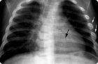
When X-rays of the ribs, the state of the bone mechanism is visualized, and the spine can be partially seen. The degree of ionizing radiation is not considered hazardous to human health, so X-rays can be considered a good alternative to ultrasound.
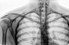
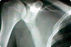
Since it is not always possible to immediately identify the cause of the pathology, doctors use an X-ray of the scapula. We are talking about a non-invasive, painless and affordable diagnostic method, which, moreover, is quite informative.
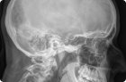
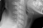
The need for an X-ray of the cervical spine is assessed by the doctor in each specific situation. In most cases, this diagnostic method allows a medical specialist to quickly and accurately determine the diagnosis and begin adequate treatment.
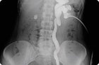
One of the X-ray methods for diagnosing diseases of the urinary system is pyelography (pyeloureterography, ureteropyelography), in which the study of the kidneys and ureters is carried out using special contrast agents.
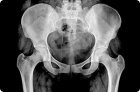
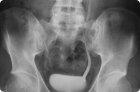
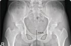
Coccyx X-ray is an informative diagnostic method that helps to identify many osteoarticular pathologies in the corresponding area of the spinal column.