Medical expert of the article

New publications
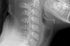
X-ray of the cervical spine with functional tests
Last reviewed: 03.07.2025

All iLive content is medically reviewed or fact checked to ensure as much factual accuracy as possible.
We have strict sourcing guidelines and only link to reputable media sites, academic research institutions and, whenever possible, medically peer reviewed studies. Note that the numbers in parentheses ([1], [2], etc.) are clickable links to these studies.
If you feel that any of our content is inaccurate, out-of-date, or otherwise questionable, please select it and press Ctrl + Enter.
The cervical spine is the most mobile part of the spine, experiencing enormous stress every day, which often leads to injuries and deformation of the vertebrae. The fact is that the muscular system in this section is somewhat weaker than in other areas of the spine. As a result, muscles, vertebrae, ligaments, and even brain tissue suffer due to the blockage of blood flow by deformed vertebrae and spasmodic muscles. Often, to determine the cause of the malfunction in the body, it is necessary to conduct an X-ray of the cervical spine - this is an accessible, accurate and fast diagnostic method for detecting basic disorders in the musculoskeletal system. [ 1 ]
Indications for the procedure
In what cases may a doctor insist on an X-ray of the cervical spine:
- for pain in the neck area, if its origin is unknown or questionable;
- in case of pain or periodic numbness in the shoulder area;
- for headaches, tinnitus of unknown origin;
- with regular crunching of the cervical vertebrae, which causes a person quite severe discomfort;
- when neck movement is limited;
- for weakness and numbness of the hands;
- in case of regular migraine attacks, dizziness, periodic deterioration of vision, constant drowsiness and apathy, impaired concentration;
- in case of injuries, falls, and other damage to the upper segment of the spinal column.
The need for an X-ray of the cervical spine is assessed by the doctor in each specific situation. In most cases, this diagnostic method allows the medical specialist to quickly and accurately determine the diagnosis and begin adequate treatment.
Preparation
A cervical X-ray is a relatively simple diagnostic procedure that does not require any special preparation on the part of the patient. There is no need to follow any special diet, take certain medications, or fast: just come to the X-ray room, take off the clothes covering the area being examined, as well as any metal objects (chain, jewelry, earrings, removable dentures). If the cervical X-ray is performed routinely, the patient should think about preparation in advance, wear clothes that are easy to remove, and leave all metal objects and jewelry at home. Why is this necessary? The structure of the metal is not able to transmit X-ray radiation, so objects made of this material will be “photographed” in the image, which may interfere with adequate examination of the image. [ 2 ]
Technique neck X-rays
X-rays of the cervical spine are usually performed with the patient either sitting or standing. In this case, the body parts that are not being examined must be covered with a special lead plate or apron (which is especially important if the diagnostics are performed on small children or a pregnant woman).
The radiologist who performs the procedure leaves the radiology room immediately at the moment the image is recorded. If for some reason his presence is necessary, he must wear appropriate lead protection.
During the recording of the image, the patient must follow the instructions of the radiologist and remain completely still. You can only move if the doctor asks you to - for example, in some situations there is a need to change your position, bend over, inhale, etc.
Sometimes the doctor insists on taking pictures in different projections, which may require repeated diagnostics.
- X-ray of the cervical spine in two projections – front and side – is a fairly common procedure that allows the doctor to examine the area being examined in more detail. To obtain a “side” image, the patient has to lie on his side – for example, on a couch. And to obtain a “front” image, he must lie on his back.
- The spinal column is particularly mobile in some places, so X-rays of the cervical spine with functional tests are often performed. Such tests require tilting or turning the head at a certain angle; sometimes the doctor asks the patient to bend, lie down, or even open his mouth. The doctor's task in this case is to select the correct angle for the X-ray tube. The patient's task is to listen attentively to the doctor and follow his instructions.
- X-rays of the cervical vertebrae are sometimes performed in combination with X-rays of other vertebral sections, such as the thoracic. In this situation, we speak of a third X-ray projection.
- X-ray of the first cervical vertebra is usually performed through the patient's open mouth. The patient lies on his back, with his arms extended along the body. The median sagittal plane of the head is placed perpendicular to the plane of the couch. The patient's head is tilted back so that the plane between the lower edge of the maxillary incisors and the lower edge of the occipital bone is perpendicular to the plane of the couch. The patient opens his mouth as much as possible, and the central beam of rays is directed vertically to the lower edge of the maxillary incisors. [ 3 ]
- An X-ray of the cervical-collar zone involves taking pictures of the back of the neck up to the level of the IV thoracic vertebra, as well as the surface of the chest up to the II rib.
- An X-ray of the cervical spine is performed on a child only if there are compelling indications - for example, in case of headaches of unknown origin, numbness in the arms, curvature of the spine, causeless pain in the arms, as well as in case of suspected tumors, pathological changes in the discs, signs of hernias and local inflammatory processes.
- An X-ray of a cervical hernia allows for excellent visualization of the problem without resorting to a more expensive MRI procedure. The hernia is accompanied by a displacement of the nucleus pulposus with a rupture of the fibrous ring: as a result, the nerve roots, a kind of spinal cord branching, are compressed. The oxygen and nutritional supply to the nerve roots is disrupted, and nerve impulse conduction deteriorates. [ 4 ], [ 5 ]
- X-rays for osteochondrosis of the cervical spine allow us to determine the level of reduction in the height of the intervertebral discs, the boundaries of the localization of dystrophic and degenerative changes in the spine, and to record marginal growths. These disorders can be observed mainly in elderly patients. Osteochondrosis causes the appearance of aching and periodic pain in the neck and/or head: if such pain is severe or constant, then additional diagnostic methods should be used in the form of computed tomography or magnetic resonance imaging. [ 6 ]
- An X-ray of a cervical vertebra subluxation is taken using a lateral projection. The image is taken in such a way that not only the cervical vertebrae are visible, but also the lower part of the occipital bone and the hard palate. The doctor determines the relationship of the upper cervical vertebrae and the size of the spinal canal using certain X-ray calculations. [ 7 ]
- X-rays for cervical instability allow us to record the displacement of the vertebrae. In fact, instability manifests itself in pathological mobility in a segment of the spinal column - for example, in an increase in the amplitude of adequate movements, or in the appearance of atypical new degrees of free mobility. Previously, it was not possible to see such a violation on an X-ray image, but now specialists have noticed that the problem is indicated by a visible displacement of the vertebrae with excessive mobility of the vertebral segments. [ 8 ], [ 9 ]
- An X-ray of cervical lordosis helps to examine the problem: an arched curve with a convexity facing forward. As a rule, this examination is carried out in case of posture disorders, pain in the cervical spine, numbness of the arms and regular headaches. Pathological cervical lordosis can occur as a result of birth trauma, diseases of the spinal column or the whole body (for example, this happens with metabolic disorders, the development of tumor processes, etc.). [ 10 ]
- X-ray of the cervical vertebrae displacement is as follows. The norm during flexion is the displacement of all cervical vertebrae relative to each other: its value is equal and does not exceed 3 mm. If this norm is exceeded, both for all vertebrae and for 1-2, against the background of the absence of symptoms of physiological mobility of the remaining vertebrae, they speak of pathological instability of the cervical spine.
- X-rays for uncovertebral arthrosis of the cervical spine allow us to see the destruction or deformation of the intervertebral discs and facet joints of the spine. Most often, the painful process occurs between the first and second cervical vertebrae. It is impossible to diagnose uncovertebral arthrosis without using X-rays of the cervical spine or MRI. [ 11 ]
Contraindications to the procedure
There are a number of contraindications, in which the doctor will not refer the patient for an X-ray of the cervical spine, choosing another, alternative diagnostic procedure. For example, X-rays are not performed:
- if the patient is in a serious and unconscious condition;
- women during pregnancy (at the discretion of the doctor);
- if the patient has an open pneumothorax.
- X-ray with contrast is contraindicated:
- in case of hypersensitivity to the composition of the contrast agent;
- in case of thyroid gland pathologies;
- in active form of tuberculosis;
- in decompensated states of liver and kidney diseases;
- in decompensated diabetes mellitus.
The period of gestation is considered a relative contraindication, and the doctor must take all necessary precautions when referring a woman for an X-ray of the cervical spine during pregnancy. However, the doctor always evaluates the possible danger of the procedure first: it is especially undesirable to do an X-ray in the first trimester and in the last trimester. If it is possible to wait with diagnosis and treatment until the birth of the baby, then this should be done, without exposing the woman and the fetus to unnecessary risk. [ 12 ], [ 13 ]
Normal performance
How does an X-ray work? Its rays, when passing through body tissues, form an image that is transmitted to a screen or reflected in a photograph. Tissues that transmit rays well will be colored in dark shades on the image, and hard tissues that absorb rays will be light.
If the examination was carried out correctly, without errors, the doctor will be able to decipher and describe the image without any problems. The description includes an assessment of the condition of the vertebrae (their height and location) and vertebral curves, the distance between the vertebrae, an analysis of darkened areas (the presence of bone integrity violations, structural changes - fractures, deformations, osteoporosis), an analysis of light areas (the presence of neoplasms or metastases, inflammatory foci). [ 14 ]
Based on the data obtained, the doctor will be able to make a diagnosis and begin appropriate treatment. If necessary, additional diagnostics will be prescribed.
Cervical ribs on x-ray
Cervical ribs are a congenital defect, mostly bilateral. Most often, the cervical ribs are diverted from the seventh cervical vertebra, less often from the sixth, fifth or fourth vertebra. Sometimes the ribs reach the sternum and are connected to it by a cartilaginous joint, or end with a free end, not reaching the sternum by about 5.5 cm. If the cervical ribs are incomplete (do not exceed 7 cm), then their ends touch the subclavian artery and the nerve plexus of the shoulder. [ 15 ]
Often, the owners of such an anomaly do not suspect its existence until an X-ray of the cervical spine is taken. However, rudiments can significantly and negatively affect a person's health: numbness, hyperesthesia, neuralgia, and finger contracture are observed in the hands. With the aggravation of neurovascular disorders, ischemic wrist contracture can develop, up to gangrene of the limb. To prevent complications, it is very important to conduct an X-ray of the cervical spine earlier, followed by surgical intervention. [ 16 ], [ 17 ]
Complications after the procedure
The radiography method is based on the action of ionizing radiation, which to a certain extent poses a risk to the patient, since it can initiate the development of oncological pathologies. It is for this reason that in medicine there is the ALARA principle, according to which the degree of influence of ionizing rays should be reduced to a reasonable minimum volume. This means that the doctor must always weigh and evaluate the potential harm from the study, as well as the danger that may occur if the X-ray is refused.
The peculiarity of X-rays is their excellent penetrating ability and the ability to affect the entire area under study. Experts explain the potential harm of the procedure by damage to the DNA of dividing cellular structures. As a result, mutations appear, which increases the risk of tumor processes. [ 18 ]
However, it is important to understand that radiation exposure is always measured and taken into account. For example, one X-ray of the cervical spine has a radiation exposure of no more than 1 mSv (millisievert). This means that such a procedure can increase a person's risk of developing cancer by approximately 0.0000055%. According to experts, the degree of such risk is significantly lower than the risk of getting into an accident while riding in a taxi or public transport.
Most doctors insist that there is no need to be afraid of radiation during an X-ray, since the risk of negative consequences is negligible, but the risk of an incorrect diagnosis or the wrong treatment is really high.
In addition, it is incorrect to correlate each occasional detection of a malignant tumor with the fact that a person underwent an X-ray of the cervical spine or another area.
In some cases, cervical X-rays need to be performed using a special contrast agent - for better visualization and clearer definition of the vascular network and tissue boundaries. Contrast is administered intravenously: as a rule, specific iodine-containing drugs are administered. But in some patients, complications may develop after such administration:
- skin rashes;
- headaches, dizziness;
- difficulty breathing;
- swelling.
To avoid them, a person is tested for allergies before the contrast agent is administered. According to medical statistics, complications of this kind occur in less than 1% of cases. [ 19 ], [ 20 ]
Care after the procedure
The patient does not need any special care after the cervical X-ray procedure. If desired, a small preventive measure can be taken to remove the minimum dose of radiation that has entered the body.
The simplest and most common preventive method is drinking a cup of milk, which does its job perfectly, binding and accelerating the removal of radionuclides. Adult patients can drink a glass of quality dry grape wine, which also neutralizes the effects of radiation.
The best substitute for dry wine is natural grape juice with pulp, or just a good big bunch of grapes, or other natural juice of your own making. Packaged juices that are sold in stores are not suitable for such purposes, and can even worsen the condition. [ 21 ]
It is advisable to add foods that contain iodine to your diet. For example, seafood, sea fish, greens, persimmons, etc. are excellent.
If the cervical spine X-ray was performed repeatedly in a relatively short period of time, then the specified products should be tightly included in the diet. In addition, it is important to regularly consume fermented milk products and vegetables.
For frequent X-ray examinations, the following products are especially recommended:
- unrefined vegetable oils;
- natural juices, decoctions and compotes from dried fruits and berries, herbal infusions;
- honey, propolis;
- rice, oatmeal;
- vegetables, greens;
- raw quail eggs.
It is important to drink plenty of fluids throughout the day after the procedure, which helps to cleanse the body faster.
Reviews
According to numerous reviews, an X-ray of the cervical spine, made with high-quality equipment, is a fairly informative diagnostic method. It is even better if the X-ray machine is digital: this will allow for a high-quality study with the lowest possible radiation dose. As experts explain, digital X-ray devices give a much lower radiation load to the body, unlike the "old" type of devices.
In addition to X-rays, magnetic resonance imaging can be used to examine the cervical spine. There is no exact answer to the question of what is better, MRI or X-ray of the cervical spine. For example, in case of traumatic injuries, a simple, accessible and quick X-ray examination is often sufficient. And MRI is prescribed in more complex or unclear cases, or when the patient is pregnant, or when it is impossible to conduct an X-ray examination for some reason. The decision to replace X-rays with a more expensive MRI procedure is made by the attending physician.