Medical expert of the article
New publications
Digital X-ray
Last reviewed: 04.07.2025

All iLive content is medically reviewed or fact checked to ensure as much factual accuracy as possible.
We have strict sourcing guidelines and only link to reputable media sites, academic research institutions and, whenever possible, medically peer reviewed studies. Note that the numbers in parentheses ([1], [2], etc.) are clickable links to these studies.
If you feel that any of our content is inaccurate, out-of-date, or otherwise questionable, please select it and press Ctrl + Enter.

What is this new diagnostic method – digital X-ray? In fact, it is a familiar X-ray examination with obtaining an image processed digitally. The digital analogue uses the latest equipment with minimal radiation exposure, which is a significant advantage. What else do you need to know about the new product? [ 1 ]
Digital or film x-ray?
First of all, most patients are interested in: what is the difference between conventional film and new digital X-ray? There are differences, and they are as follows:
- the digital image is displayed not on film, but on a computer screen, and then, if necessary, transferred to a disk or other storage device;
- the entire process of scanning and displaying results takes no more than ten minutes;
- the image is of the highest quality;
- the image can be further processed using various programs – for example, to improve the visualization of a specific area;
- the dose of radiation received during the procedure is less than during a conventional X-ray examination;
- the diagnostic results can be immediately sent to the attending physician’s computer;
- Digital x-rays are safe and can be stored for a long time.
Radiation exposure in digital x-ray
The issue of radiation dose during X-ray examinations has always been quite relevant. Experts have calculated that when performing digital X-rays, the radiation load is approximately ten times less than during a conventional film examination. This is especially important if the diagnostics are prescribed to a child or a woman during pregnancy or breastfeeding.
It is important to understand that the higher the quality and the newer the device used to obtain the X-ray image, the more accurate and safe the examination will be. If you aim to minimize the adverse effects of the procedure on the body, try to choose a clinic that has the most modern equipment. [ 2 ]
Indications for the procedure
Digital X-ray has many indications, just like its film counterpart. The examination is prescribed:
- in case of lung diseases, or if they are suspected, as well as for preventive purposes for the timely detection of dangerous pathologies;
- for diagnostics of cardiovascular diseases, heart defects, functional disorders of the pulmonary circulation;
- for the diagnosis of fractures, curvatures and other pathologies of the spinal column, including osteochondrosis;
- for diseases of the stomach and duodenum - with or without contrast;
- to assess the functioning of the biliary system (usually performed with contrast);
- to detect polyps, tumor processes, foreign bodies, inflammatory reactions in the large intestine;
- for diseases of the abdominal cavity, which are accompanied by severe abdominal pain;
- for diseases of the musculoskeletal system - for example, fractures, dislocations, ligament damage, chronic joint problems;
- in dentistry before and after dental treatment, during implant placement, in case of abscesses, jaw fractures, and bite disorders.
Preparation
If the patient is to undergo a digital X-ray of the extremities, chest, cervical or thoracic spine, then no special preparation is required for the procedure. However, if it is necessary to obtain an image of the lumbar or sacral spine or abdominal organs, then some preparation rules still exist. For example, a few days before the examination, it is necessary to change the diet, excluding foods that cause increased gas formation: peas, beans, whole milk, baked goods, soda. If there is a tendency to flatulence, then three or four days before the procedure, you can take enzyme preparations that promote digestion. Such measures are necessary to reduce the amount of gases in the intestines, since they can negatively affect the clarity of the X-ray image, as well as complicate its interpretation. [ 3 ]
Before the digital X-ray procedure, you must not drink alcohol or smoke. Immediately before entering the X-ray diagnostic room, you must remove all metal objects (jewelry, watches, etc.), remove your mobile phone, keys, etc. from your pockets.
The device for carrying out the procedure
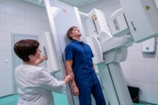
Digital X-ray machines can be either mobile (portable) or stationary. The most functional is the digital X-ray system, which can be used for any type of X-ray. We are talking about a universal diagnostic complex, suitable for both conventional fluorographic screening and for specific X-ray examinations of the extremities, abdominal or thoracic organs, spinal column, skeletal system (including facial and skull bones). [ 4 ]
Modern digital X-ray machines are convenient and safe for both the doctor and the patient. The resulting image is of high quality due to the increased output power and short exposure period. The information obtained during the procedure is easily integrated into the hospital-wide network.
Technique digital X-ray
To obtain a high-quality digital image, the patient must adhere to the following algorithm of actions:
- take the position of the body and limbs recommended by the radiologist and do not move until the end of the procedure;
- It is advisable to hold your breath from the moment the device is turned on: this is necessary if an X-ray of the lungs or thoracic spine, as well as the lumbar region and abdominal organs is being performed.
The result is interpreted by a specialist immediately after the procedure; the patient's participation in this process is not required. The radiologist examines the resulting image, evaluates pathological changes and makes a conclusion. For most patients, the transcript is given in person some time after the end of the study, but it is possible to transfer the information directly to the attending physician's computer. [ 5 ]
After the digital X-ray procedure, the patient can go home or to the hospital, depending on the situation. If the patient cannot move independently, he is transported by accompanying persons - medical workers or relatives.
Digital chest x-ray
A digital chest X-ray may be recommended by a doctor for various reasons – both to make a diagnosis and to assess the dynamics of a disease or for preventive purposes during routine examinations. Most often, the procedure is prescribed:
- in case of pneumonia;
- pleurisy, bronchitis;
- tumor processes in the lungs;
- for tuberculosis, etc.
If a patient goes to a doctor and complains of a prolonged cough, difficulty breathing, chest pain, a feeling of heaviness and wheezing, then X-ray diagnostics will be recommended. Standard preventive fluorography can also be performed digitally, which is safer and faster.
Digital X-ray is especially recommended for pregnant women, children, medical workers, military personnel, patients who have had tuberculosis, patients with chronic respiratory pathologies, as well as all those who for one reason or another are forced to undergo frequent X-ray examinations. Using a digital analogue will significantly reduce the overall radiation load on the body.
Digital chest x-ray
Chest X-ray is always prescribed for strict indications. For example, the procedure is indicated if the patient complains of difficulty breathing, hemoptysis, chest pain, if there is an injury to a difficult part (spine, sternum, collarbones or ribs). Diagnostics are done if pneumonia or malignant tumors are suspected.
What does a chest x-ray show:
- pneumonia;
- tuberculosis;
- pulmonary emphysema;
- malignant neoplasms;
- chest trauma, foreign bodies in the respiratory system;
- cardiac tamponade, pericardial effusion.
To interpret the results, the specialist will analyze the dark spots and shadows, and the accuracy of the image will depend on how clearly the instructions were followed during the examination, as well as how correctly the projection was selected and set up. [ 6 ]
When evaluating a digital image, the doctor must take into account the tissue structure, size and shape of the lungs, features of the lung fields, and localization of the mediastinal organs. Dark spots may indicate an inflammatory process, and light spots on the lung picture indicate a parenchyma disorder with the formation of abscesses, caverns, etc.
Digital X-ray of the spine
Performing a digital X-ray of the spine has some peculiarities. The examination itself is not complicated, it is safe and lasts no more than 10-20 minutes. Before the procedure, the patient must take off his clothes (most often they undress to the waist, unless it is necessary to diagnose the coccyx area).
The neck and lumbar region are the most mobile segments of the spine, so when examining them, it is important to use functional tests. The radiologist may ask the patient to tilt or turn his head, bend or straighten, lie down, raise his arms, etc. It is very important to give the spine the necessary position so that the required zone is most “open” for visualization.
The sacrum, coccyx and thoracic region are not so mobile, so they are photographed using two projections. The patient can sit or lie down: the best body position will be suggested by a radiologist.
Patients with spinal injuries are transported on stretchers for digital X-rays.
Digital x-ray of the stomach with barium
Digital X-ray of the stomach is a type of abdominal fluoroscopy that helps to examine organ pathologies. Ulcers, polyps, dystrophic and inflammatory processes, and oncological neoplasms are “in the crosshairs”. During the procedure, the doctor can also pay attention to organs located in close proximity: the esophagus and duodenum.
Before prescribing a digital X-ray to a patient, the doctor must rule out the possibility of the patient developing an allergy to the contrast component. If everything is in order, the patient is recommended to follow a special diet for three days.
When performing an X-ray, the patient drinks two sips of a special substance (barium), after which the specialist records the image of the esophageal walls. Then the patient drinks another 200 ml of contrast agent, and the radiologist records the image of the stomach.
The entire procedure usually lasts about half an hour. If you need to visualize the duodenum, you need to wait a little longer until the barium gets into the cavity of the organ.
The images can be taken from different angles: the patient lies on the couch on his side, on his back, on his stomach, or stands vertically. To diagnose a hernia of the esophageal orifice, the patient lies down and lifts his pelvis at an angle of about 40°.
For the patient, digital X-ray with barium is not dangerous: the substance completely leaves the stomach in about 60-90 minutes. Sometimes constipation occurs after the diagnosis, the color of the feces changes. As a rule, the process of defecation returns to normal on its own within a couple of days.
For reference: the contrast agent is barium sulfate diluted with drinking water. The substance tastes like a calcium solution (chalk) and is usually well accepted by patients. The examination is not accompanied by discomfort, and the information obtained allows you to quickly identify serious problems that are difficult to visualize using other diagnostic methods.
Digital X-ray for a child
Digital X-rays in children can be performed even from birth, if there are appropriate indications for this. With this method, it is possible to examine and assess the condition of internal organs, the musculoskeletal system - in a word, almost all tissues of the body:
- X-ray examination of the brain will allow visualization of the presence and condition of metastases, the shape of the cranial bones, the quality of the vascular pattern, the condition of the paranasal sinuses and cranial sutures;
- When performing digital X-rays of the lungs, it is possible to identify tumor processes, pneumonia, bronchitis and fibrosis;
- X-ray of the abdominal area helps to identify neoplasms, metastases, abscesses and foci of destruction in tissues;
- The procedure of digital X-ray of the spine is performed in case of injuries, as well as to exclude hernia, infectious lesions and oncological diseases.
When performing diagnostics on children, it is very important to ensure that the child remains completely still for a few seconds or minutes. Many clinics for babies have a special X-ray "cradle" in which the baby is fixed in the required position. In rare cases, if it is impossible to hold the baby, short-term anesthesia may be used.
It is strictly forbidden to use digital X-ray on a child on your own: the examination is performed only upon referral from a doctor. The doctor evaluates the need for using the method after an external examination, collection of anamnesis and preliminary laboratory diagnostics. [ 7 ]
Contraindications to the procedure
Digital X-rays have relatively few contraindications, and none of them are categorical or too strict. For example, if the examination is prescribed to a pregnant woman, it is better not to conduct it during the first trimester. However, if there are indications, X-rays are still done, but all necessary protective equipment is used.
The lactation period is also considered a relative contraindication. However, even here the procedure is performed in the presence of injuries and diseases, for the diagnosis of which it is impossible to do without an X-ray.
The situation is more complicated if the patient suffers from hypermobility – for example, such a condition is typical for schizophrenia, some psychoses and neuroses. Due to the fact that a person cannot ensure immobility for some time, the procedure may be at risk, because the resulting images will be blurred and unclear.
Complications after the procedure
When performing a digital X-ray, the patient receives a relatively small dose of radiation, which on average corresponds to 4-6% of the annual norm received by a person from natural radiation sources (this norm is defined as approximately 3 mSv per year). That is, approximately the same amount of radiation can be received by sunbathing for an hour in the sun. And yet, to avoid unpleasant complications, doctors do not recommend performing digital X-rays too often - that is, more than six or seven times a year.
It should not be forgotten that X-ray diagnostics are prescribed according to strict indications, and often the goal of doctors is to identify a life-threatening pathology. If saving not only health but also a person's life is at stake, then possible or impossible complications after an X-ray are usually not discussed.
Digital X-ray is the best option among those currently available, as it is safer and no less informative than the X-ray examination we are used to. If possible, during the procedure, it is necessary to provide protection for the organs that will not be examined: for example, special plates are put on the chest and abdomen area that do not allow dangerous rays to pass through.
Consequences after the procedure
The impact of radiation on the human body may depend on both the duration of the procedure and its quality: the newer and more modern the digital X-ray equipment, the safer the diagnostics. The unit of measurement of radiation dose is Sievert. Each X-ray room has special dosimeters that measure the degree of human radiation during the examination.
The radiation dose is directly related to the quality of the equipment. Thus, digital X-rays are accompanied by a lower degree of radiation than the usual film analogue. It should also be taken into account that a higher dose of rays is used to obtain an image of the skeletal system than to examine hollow organs.
Many patients are afraid of X-ray diagnostics because of the high probability of negative health consequences. On the one hand, any amount of radiation can harm the body. And, on the other hand, the possible danger that exists when refusing an X-ray significantly exceeds this harm, as it can lead to serious complications from the damaged organ or system. Therefore, if there are indications for the study, it should still be done. Of course, for greater safety, it is better to choose digital X-ray: modern devices give a much lower radiation load on the body. [ 8 ]
Care after the procedure
Special care after digital X-ray is usually not required, but doctors have identified several recommendations to speed up the removal of the received radiation dose from the body:
- upon arrival home you should immediately take a warm shower;
- You need to drink a lot of water during the day.
Other drinks also speed up the cleansing of the body:
- green tea;
- fresh milk;
- natural juice with pulp (peach, apple, strawberry, etc.);
- grape juice.
In addition, it is recommended to walk a lot in the fresh air, preferably in the shade - for example, in a forest or park. Sunbathing and staying in the sun for a long time on the day of the study is not advisable. [ 9 ]
Modern digital X-rays allow obtaining clear and high-quality images, thanks to which the doctor gets the opportunity to adequately assess the clinical features of the problem and choose the optimal treatment tactics. Today, such a study can be done in many clinical centers: information about the type of digital device and its capabilities is provided directly in the specific selected clinic.

