Medical expert of the article

New publications
X-ray of the coccyx
Last reviewed: 03.07.2025

All iLive content is medically reviewed or fact checked to ensure as much factual accuracy as possible.
We have strict sourcing guidelines and only link to reputable media sites, academic research institutions and, whenever possible, medically peer reviewed studies. Note that the numbers in parentheses ([1], [2], etc.) are clickable links to these studies.
If you feel that any of our content is inaccurate, out-of-date, or otherwise questionable, please select it and press Ctrl + Enter.
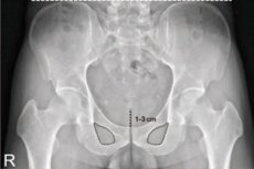
Such a diagnostic examination as a coccyx X-ray is not prescribed very often, but only if the patient complains of discomfort or pain in this area of the spine. The examination itself is simple and can be performed in almost any outpatient facility that has X-ray equipment. A coccyx X-ray is an informative diagnostic method that helps to determine many bone and joint pathologies in the corresponding area of the spinal column.
Indications for the procedure
X-ray is a common diagnostic method based on the use of gamma rays. The method is so popular and accessible that it is used in almost any clinic and private medical and diagnostic institutions. And this is not surprising, because X-ray has a number of advantages, such as effectiveness (information content), ease of implementation and affordability.
X-ray of the coccyx is prescribed in case of suspected injury of the specified area or inflammatory process. The following list of indications for this examination can be distinguished:
- severe bleeding in the coccyx area;
- pain, pressure, or numbness in the tailbone area; [ 1 ]
- visible vertebral displacement in the lower back;
- suspicion of dislocation, subluxation or fracture of the coccyx;
- diseases of the pelvic organs;
- urinary or defecation disorders;
- limited range of motion in the lower back;
- a feeling of stiffness in the lower limbs;
- suspicion of intervertebral hernia;
- inflammatory diseases of the lower spine;
- suspicion of oncopathology.
A coccyx X-ray for preventive purposes can only be prescribed to patients who, due to their professional or other characteristics, are forced to spend a long time in a sitting position. The examination is mandatory for people who have fallen from a height onto their feet or lower back.
X-ray of the coccyx is also relevant for degenerative processes in this area.
Preparation
X-ray of the coccyx is a technically simple procedure. However, it is necessary to prepare for it in advance - except in emergency cases, when patients are taken to the emergency room with severe injuries.
When performing a planned coccyx X-ray, it is better to prepare for the examination in advance. This is necessary to ensure that reliable information is obtained on the image.
Preparation consists of the following stages:
- 2-3 days before the coccyx X-ray, you should review your diet and exclude heavy (fatty, hard to digest) dishes, as well as foods that can cause fermentation and increased gas formation in the intestines. It is advisable to temporarily give up peas, white cabbage, sweets and yeast pastries, mineral water, dried fruits, whole milk. It is important not to overeat during these days.
- The day before the examination, the intestines are cleaned using an enema. You can use clean warm water or chamomile infusion. In case of strong accumulations of feces, it is additionally allowed to take a laxative (for example, Duphalac).
- On the eve of the procedure, you are allowed to have a light snack. But it is better to go to the diagnostics with an empty stomach.
When going to the X-ray room, you should leave all metal accessories and jewelry at home. It is advisable to wear comfortable clothes that can be easily removed and put on just as easily and quickly. If there are non-removable metal implants that may interfere with the examination, it is better to immediately inform the doctor about them.
Enema before coccyx x-ray
Cleansing the intestines before taking a coccyx X-ray is an important measure to ensure the quality of the image. Of course, you can take an image without first giving an enema, but in this situation there will be a risk of some image distortion.
An enema is not performed before an X-ray:
- in emergency cases when the patient's condition is serious;
- in case of fever, high body temperature;
- for diarrhea;
- for inflammatory bowel diseases (including the rectum);
- for abdominal pain, nausea of unknown origin;
- after a recent heart attack or stroke;
- in case of internal bleeding.
If an enema is still indicated, then it is best to perform it using an Esmarch mug - a capacious vessel that rises to a certain height, creating optimal water pressure.
Cleansing the intestines with an enema is done with warm clean water or herbal infusion (chamomile, calendula). The volume of liquid used is directly proportional to body weight: the greater the weight, the greater the volume of liquid used. As a rule, 1 to 2 liters of water is enough for one enema for an adult. [ 2 ]
Here's how to administer an enema:
- the enema system is filled with liquid;
- clamp the tube and hang the container with water at the optimal height;
- prepare a place for the procedure - for example, lay out an oilcloth;
- apply a little Vaseline or vegetable oil to the tip;
- the patient lies on his side or takes a knee-elbow position, after which the enema tip is inserted into the rectum (approximately 8-10 cm), the clamp is released and the required volume of liquid is gradually poured in;
- then the tip is removed;
- The patient should, if possible, retain the liquid in the intestines for at least five minutes.
If at any stage the patient’s condition worsens, pain appears, etc., then the procedure is stopped.
To perform a colon cleansing enema more comfortably, you should pay attention to the following points:
- if pain occurs during the introduction of water into the intestine, it is necessary to reduce the rate of delivery;
- do not use cold or hot water for enemas (optimally from +27 to +38°C);
- Be sure to remember to lubricate the tip with Vaseline or vegetable oil;
- It is important to perform the procedure smoothly, without haste, calmly.
If there are any problems with administering an enema before an X-ray of the coccyx, or if there are contraindications, you can consult a doctor: in some cases, it is allowed to cleanse the intestines using laxatives or microclysters (Microlax, etc.).
Technique a coccyx X-ray
X-ray of the coccyx usually covers the sacrococcygeal area. The examination is performed in a horizontal position: the patient is placed on a special couch (table). It is possible to obtain images from different positions or body positions, which is discussed with the doctor in advance:
- X-ray of the coccyx in the direct posterior projection is performed as follows. The patient lies on his back, bends his legs at the knee and hip joints (or only at the knees). The arms are extended along the body.
- X-ray of the coccyx in the lateral projection is performed from the side position. The patient raises the upper limbs and places them behind the head. The lower limbs are at a slight angle.
- Oblique projection is used rarely and only to clarify the functionality of a particular joint.
During the X-ray, the doctor may take one or two pictures. If there are difficulties in visualizing individual segments and joints, the doctor may refer the patient for additional diagnostics, such as MRI or CT. [ 3 ]
How is an X-ray of the coccyx done?
Immediately before the coccyx X-ray, the patient enters the office, removes all metal objects and accessories (watches, chains, piercing jewelry, etc.), and removes clothing that may interfere with obtaining an image of the required area of the body.
The patient then lies down on a special table or couch near the X-ray machine in such a way that the scanning device is located above the lower back. If necessary, the radiologist will correct the position and give appropriate recommendations.
During the procedure, images are taken in the required projection. If there are several such projections, the doctor will inform the patient about the need to change the position of the body.
As a rule, the entire diagnostic session for an X-ray of the coccyx takes no more than 15 minutes.
After the procedure, the radiologist develops the film, examines the image, writes a description and gives the results to the patient or sends them to the attending physician who previously issued the patient a referral. In turn, the attending physician, based on the results of the diagnostics, establishes a final diagnosis and prescribes the appropriate treatment. [ 4 ]
What does a coccyx x-ray show?
X-ray of the coccyx helps to examine traumatic injuries or inflammatory processes. Most often, it is used to diagnose the following pathologies:
- A coccyx hematoma is a blood leak into the tissue due to a bruise. Hemorrhage in this area usually does not resolve, so minor surgery may be required, primarily to prevent the development of an inflammatory process with suppuration. In this case, an X-ray of the coccyx helps the doctor assess the severity of the injury and the likelihood of complications.
- Sacral dislocation is a pathological deformation caused by a coccyx injury. The main signs of the pathology are considered to be a pronounced external displacement, pain when palpating, swelling and clicks (crunching) when trying to move.
- A coccyx fracture is one of the most complex traumatic pathologies, which can be both open and closed. An open fracture is accompanied by severe pain, and a closed fracture is accompanied by symptoms of varying intensity, depending on the complexity of the injury. Often, an X-ray of the coccyx does not allow for a thorough examination of the problem, so the doctor additionally prescribes a CT scan.
During an X-ray of the coccyx, other pathologies can also be identified, in particular, tumors, osteochondrosis, inflammatory processes, hernial protrusions, etc. [ 5 ]
X-ray of tailbone fracture
A coccyx fracture can be the result of a fall on a hard surface (asphalt, tiles, ice), or the result of a direct blow. Women sometimes get a coccyx injury during childbirth. It is believed that female patients suffer from such injuries more often – primarily due to the greater width of the hips. It turns out that the coccyx is more vulnerable in women.
A tailbone fracture is accompanied by severe pain: it is almost impossible to sit. Hematomas may form in the lower back, and pain bothers, including during defecation. With minor damage, painful sensations are noted during sexual intercourse.
A coccyx fracture is often combined with a dislocation (called a "fracture-dislocation"), with the displacement of fragments depending on the direction of the damaging force relative to the axis of the coccyx, which becomes visible on an X-ray. When displaced, muscles and ligaments are usually damaged.
X-ray of a bruised tailbone
It often happens that people fall and damage the lowest segment of the spine – the coccyx. This area is a series of interconnected vertebrae, which in our ancestors was nothing more than a part of the tail. Experts consider the coccyx to be one of the most vulnerable parts of the spine. That is why bruises of this area are often diagnosed in patients of any age, and especially in winter – on ice.
A small bruise of the coccyx as such is not displayed on an X-ray. The examination is carried out in order to exclude more complex injuries of the vertebral segment - in particular, a fracture (which, by the way, happens relatively rarely) or dislocation. The scale of treatment will depend on the results of the X-ray of the coccyx.
Angular deformity of the coccyx on x-ray
A severe contusion of the coccyx may be accompanied by a sharp deviation of it to the side directly opposite to the direction of the traumatic factor. In this case, the sacrococcygeal ligaments remain intact. A milder traumatic impact is accompanied by the return of the coccygeal vertebrae immediately after the end of the damaging force.
If a large hematoma forms in the area of injury, it can compress the surrounding tissues and nerve endings, which entails not only pain, but also angular deformation of the coccyx. If scar tissue forms in the specified area, such deformation becomes protracted (chronic), which is clearly visible on an X-ray.
In case of partial damage to the sacrococcygeal ligamentous apparatus against the background of a hematoma, the joint of the same name may become destabilized, as a result of which its mobility increases.
Contraindications to the procedure
In case of emergency, X-ray of the coccyx is prescribed practically without restrictions. However, possible risks still exist. For example, the study is not recommended:
- during pregnancy and breastfeeding;
- in case of diagnosed tuberculosis (regardless of the stage of the process);
- patients with mental disorders;
- patients with severe obesity.
Relative contraindications may include early childhood, acute cardiovascular diseases, and thyroid disease.
It is not recommended to take X-rays too often (optimally 1-2 times a year). [ 6 ]
What is better? X-ray or MRI of the coccyx?
Patients who are faced with the need to assess the condition of the coccyx often wonder: what is better, more informative and safer - X-ray or magnetic resonance imaging? Indeed, these diagnostic methods are very different, and each patient needs to be aware of this.
MRI is used to diagnose various pathologies in tissues. Thanks to MRI, it is possible to assess the condition of internal organs, soft tissue and vascular structures, to determine benign or malignant tumor processes. The main difference between MRI and X-ray is the ability to obtain images of organs in the required plane and with a three-dimensional image.
X-rays are prescribed to detect disorders of the musculoskeletal system, so this examination is more appropriate for coccyx injuries. In addition, X-ray diagnostics are more accessible and have a low cost compared to expensive MRI. [ 7 ]
If we talk about harm, then X-ray radiation is certainly more harmful - if it is carried out frequently, or if many images are taken simultaneously in different projections. But MRI is not done to patients with claustrophobia and fixed metal implants. Considering all the pros and cons, we can say the following: if it is necessary to diagnose an uncomplicated injury and take only 1-2 images, then it is more appropriate to take an X-ray of the coccyx. MRI is indicated for complex diseases involving soft tissues and blood vessels.