Medical expert of the article

New publications
X-rays of the ribs
Last reviewed: 06.07.2025

All iLive content is medically reviewed or fact checked to ensure as much factual accuracy as possible.
We have strict sourcing guidelines and only link to reputable media sites, academic research institutions and, whenever possible, medically peer reviewed studies. Note that the numbers in parentheses ([1], [2], etc.) are clickable links to these studies.
If you feel that any of our content is inaccurate, out-of-date, or otherwise questionable, please select it and press Ctrl + Enter.
Among the numerous diagnostic studies, rib X-rays are one of the most common. Most often, the procedure is prescribed when a rib fracture is suspected. If multiple injuries are detected, the doctor may insist on conducting a survey X-ray, which is necessary to obtain more objective and complete information about the damage. A survey X-ray shows existing damage to internal organs and the chest as a whole.
When X-raying the ribs, the condition of the bone mechanism is visualized, and the spine can also be partially seen. The degree of ionizing radiation is not considered dangerous to human health, so X-rays can be considered a good alternative to ultrasound, [ 1 ] computed tomography and magnetic resonance imaging. [ 2 ]
Indications for the procedure
The thoracic skeletal frame is a reliable protection of internal organs. An X-ray of the ribs is, in fact, the same X-ray of the chest, during which it is possible to examine not only bone structures, but also the heart, lungs, respiratory tract, and spinal column. During the examination, the doctor can see damage or a violation of the shape of the bones, or the development of some pathological process.
An X-ray of the ribs is necessary if the specialist suspects the presence of such diseases and conditions:
- traumatic chest injuries;
- violation of the integrity of the ribs;
- tumor processes in the chest organs;
- foreign bodies in the chest area;
- pulmonary pathologies;
- bone tuberculosis;
- impaired bone formation, rickets;
- diseases of the spinal column;
- diaphragmatic hernias.
X-rays of the ribs are often prescribed not only as part of the diagnosis of the disease, but also to study the dynamics of the pathology and determine treatment tactics. [ 3 ]
Preparation
There is practically no need for preliminary preparation for the patient. A day before the planned examination, it is advisable to exclude products that cause increased gas formation in the intestines (peas, white cabbage, carbonated drinks), since excess gases will lift the diaphragm, putting pressure on the lungs and ribs.
Immediately before the X-ray, the patient is asked to remove their outer clothing and undress to the waist. If there are any decorations in the neck or chest area, they must be removed. If a person has long hair, it must be pulled up: it must not fall into the image area.
Before the examination, the patient should inform the radiologist about previous pathologies, surgical interventions on the chest organs, the presence of foreign objects, implants in the area being examined. Women must inform about pregnancy.
It is recommended to take all medical documents that the doctor may need: results of previously conducted examinations, established diagnoses, sheets with prescribed treatment, etc. All this can help the specialist deciphering the radiograph to issue a more informative conclusion. [ 4 ]
Technique rib X-rays
In most cases, rib x-rays are performed in direct and lateral projection. This approach allows assessing the general condition of the chest. If we are talking about a certain area of the chest, then targeted x-rays of the affected ribs are performed.
The patient undresses to the waist, presses his chest to the screen and inhales deeply (so that the chest expands), holding his breath. At the moment of expansion of the intercostal spaces, the costal contours become more distinct: this is when the specialist takes the picture.
The patient's positioning during rib X-ray may vary, depending on the area being examined and the nature of the pathology. For example, when performing a direct posterior image of the lower ribs, the person is placed horizontally on his back. In this case, the midclavicular line of the side being diagnosed should be located along the mid-longitudinal line of the couch. The upper limb is extended along the body, the legs are bent at the knees. Along the frontal plane, the body should be parallel to the plane of the couch. This position allows for a good view of the lower ribs, especially against the background of intense darkening of the liver. [ 5 ]
If it is necessary to perform a direct anterior rib x-ray, the patient is placed on his stomach, a small elevation is placed under his head and the face is turned to the side opposite to the diagnostic side. The arms should be extended along the body, the forearm and the back of the hand should be adjacent to the table.
When taking a lateral rib image, the patient is placed on the side being diagnosed, with the upper limbs raised and placed behind the head. The frontal plane of the body is parallel, and the sagittal plane is perpendicular to the plane of the couch.
To obtain an anterior oblique image, necessary for studying the condition of the anterolateral costal sections, the person is placed on the abdomen. The half of the chest being diagnosed should be tightly adjacent to the surface of the couch, and the opposite half should be slightly raised. The frontal plane of the body should intersect the plane of the couch at an angle of 40-45 degrees. The upper limb on the side of the examination is extended along the body, with the back surface adjacent to the couch. The other arm is bent at the elbow, the palm rests on the table. The criterion for adequate positioning is obtaining a clear image of the anterolateral sections of the ribs. [ 6 ]
To obtain a posterior oblique image, necessary for studying the condition of the posterolateral sections of the ribs, the patient is placed horizontally on his back, turning along the longitudinal axis of the body to the right or left (depending on which side needs to be examined), until the angle in the area of intersection of the frontal plane of the body and the plane of the couch reaches 40-45 degrees. Elevations can be placed under the back, pelvis, thigh and knee. The upper limb on the side being examined is extended along the body, and the other is moved back, resting on the edge of the couch.
In addition to a general image in different projections, sometimes it is necessary to conduct a targeted X-ray. For this, they try to bring the part of the rib with suspected pathology to the central or edge-forming position.
Contraindications to the procedure
It is important to remember that there are contraindications to performing rib x-rays:
- the first trimester of pregnancy (or the entire period of pregnancy, depending on the situation);
- severe condition of the patient, various decompensated conditions;
- open pneumothorax, bleeding;
- mental disorders, inadequacy in behavior;
- sometimes – the patient is obese.
Most experts point out that there are no absolute contraindications to performing rib x-rays, and for such categories of patients as pregnant women and children, the examination should be carried out only in the presence of strict indications and when it is impossible to use other alternative diagnostic methods. [ 7 ], [ 8 ]
Normal performance
The structural elements that limit the thoracic cavity are the rib cage, soft tissues, and diaphragm. The boundaries of the thoracic cavity are:
- ventral border – sternal segments;
- dorsal border – vertebral bodies and ribs;
- lateral borders – ribs, intercostal soft tissue, subcutaneous structure;
- caudal border – diaphragm.
The cranial thoracic region is limited by the soft tissue of the ventral cervical region and the entrance to the thoracic cavity.
During the diagnostics of the above structures and organs, it is important to clearly assess the location of the pathological process. If necessary, additional X-rays should be taken from other projections.
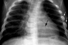
An X-ray of a broken rib shows the presence of objective signs - in particular, the fracture line, which is lighter in the image than the bone. It is also possible to change the bone structure, displacement of fragments. An indirect symptom may be a change in the adjacent soft tissues, which is also well visualized in the image - this is a darkening on the rib on the X-ray, the disappearance of physiological enlightenment in the area of the joints, thickening and compaction of the shadow of the soft tissues, which is due to the formation of hematomas and edema. [ 9 ]
An X-ray of a rib fracture does not always show specific signs, so the doctor often has to prescribe a CT scan for the patient.
A disorder such as Lyushko's rib is an abnormal development of the rib cartilages, in which their anterior section is split. The disorder is predominantly one-sided, but it cannot be called a pathology, since it is not complicated by anything and does not affect a person's quality of life. [ 10 ]
On X-ray, the Lushko rib appears as a dense formation, bifurcated in the anterior part, usually localized near the sternum. The defect is detected quite rarely (about 1% of cases).
Chondroma is a benign tumor that forms on the basis of mature cartilaginous tissue (mainly hyaline cartilage). The neoplasm grows and develops slowly, and is asymptomatic for a long time. The first signs begin to bother with compression of surrounding tissues, with spread to the pleura and damage to nerve fibers. In such situations, deformation of the chest and the appearance of costal pain are noted. Chondroma located on skeletal bones can be determined using a regular X-ray. For example, if such a tumor is localized on the costal arch, then the X-ray can see the focus of dysplasia and the cystic neoplasm itself. Rib chondroma is not noticeable on an X-ray against the background of soft tissues, since it is not radiopaque. Therefore, for other tumor localizations, diagnostic methods such as computed tomography, magnetic resonance imaging, as well as biopsy and examination of a micropreparation are used. [ 11 ]
Another congenital pathology – cervical ribs – is characterized by the presence of additional ribs in the cervical segment of the spinal column. Cervical ribs on an X-ray look like bone plates symmetrically located on the sides of the spine. Normally, they are absent, and their detection allows us to talk about a developmental anomaly. Less often, such cervical elements are located only on one side.
Numbering of ribs on x-ray
The ribs are numbered from top to bottom: as they approach the pelvic area, these bones become softer and thinner.
The first rib is located near the collarbone, and the tenth is slightly below the xiphoid process. The body of the first seven costal pairs has a gradual transition into cartilage tissue, then they connect to the rib cage.
The first and strongest seven pairs of ribs are called true, and the eighth, ninth and tenth pairs are called false ribs, since they have a cartilaginous connection between them. The eleventh and twelfth pairs are mobile, free, and are attached only on one side - to the spine.
The skeleton of an adult normally includes twelve pairs of ribs. It happens that during development a child develops a thirteenth pair, localized at the level of the seventh or eighth cervical vertebra. Another rare anomaly is the formation of one rudimentary rib in the cervical region.
Complications after the procedure
There is a certain danger in conducting an X-ray of the ribs during pregnancy. The greatest probability of complications occurs in the first trimester - that is, the first 12 weeks of gestation. It is during this period that the formation of the future vital systems of the future baby occurs. [ 12 ] Therefore, exposure of the mother's body to a large number of X-rays can have various adverse effects, depending on the period at which the study is conducted:
- first 2 weeks of gestation: embryonic death, spontaneous abortion, ectopic implantation;
- 3-4 weeks: early fetal development disorders, spontaneous abortion;
- 5-6 weeks: abnormal development of the baby’s glandular system, disturbances in the formation of the immune, nervous, and hematopoietic systems;
- Week 7: development of disorders of the digestive system and metabolic processes;
- 8th week: pathologies of the musculoskeletal system, formation of the oral cavity;
- Week 9: development of respiratory and reproductive system disorders;
- 10-11 weeks: heart defects, dental problems;
- Week 12: problems with the development of immunity and the functioning of the baby’s thyroid gland.
After 12 weeks, the negative impact of radiation on the fetus decreases. However, doctors strongly advise against x-rays for women without compelling indications. If possible, it is better to wait until the end of the pregnancy period and only then conduct diagnostics. [ 13 ]
If there is a rib injury or other problem for which other diagnostic methods cannot be used, and an X-ray is indispensable, then the examination is carried out in accordance with the following recommendations:
- women's pelvic and abdominal areas are covered with protective aprons and pads;
- inform the expectant mother about possible consequences and complications.
Consequences after the procedure
The occurrence of negative consequences or complications after the rib x-ray procedure is extremely rare. If the patient has no contraindications to the examination, and the x-ray itself is performed correctly, using special protective equipment, then adverse consequences can occur with an extremely low probability.
In general, the likelihood of side effects depends on the individual radiosensitivity of the patient, the amount and duration of radiation received. Theoretically, reactions are possible:
- from the nervous system (increased irritability, headache, dizziness, sleep disturbance);
- from the digestive tract (nausea, vomiting, diarrhea, changes in appetite, dry mouth, the appearance of an unpleasant taste in the mouth);
- from the hematopoietic system (decreased levels of neutrophils and lymphocytes, monocytes, rarely - minor eosinophilia, thrombocytopenia).
Women are more at risk of complications during pregnancy.
Care after the procedure
There are no strict restrictions or specific recommendations regarding care and regimen after an X-ray of the ribs. Some restrictions may be directly related to the injury or pathology for which the X-ray was prescribed.
Some patients worry about the radiation exposure that the patient's body receives during the diagnostic procedure. The most important thing for accelerating the removal of radioactive substances from the body is to adhere to a drinking regimen with at least 2 liters of water per day. In addition to water, you are allowed to drink freshly squeezed juices, fruit drinks, and green tea. It is allowed to drink a little dry red wine. Grapes, pomegranates, nuts, and dairy products also have anti-radiation properties. Prunes, flaxseed, and nettle leaves are useful. It is advisable to include sour cream, cottage cheese, carrots, beets, buckwheat, and seafood in your diet. But you should avoid products with preservatives, dyes, flavorings, and flavor additives, as well as smoked foods and semi-finished products.
X-rays of the ribs are safe, although they are only performed if indicated, since they involve a certain degree of radiation. The examination is prescribed only if other diagnostic methods cannot provide sufficient information about the patient's condition. X-rays should not be frightening: if you follow all the doctor's recommendations, the diagnostics will not cause the development of negative manifestations and consequences in the body.