Medical expert of the article

New publications
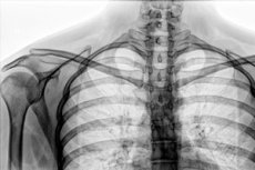
X-ray of the clavicle in two projections
Last reviewed: 03.07.2025

All iLive content is medically reviewed or fact checked to ensure as much factual accuracy as possible.
We have strict sourcing guidelines and only link to reputable media sites, academic research institutions and, whenever possible, medically peer reviewed studies. Note that the numbers in parentheses ([1], [2], etc.) are clickable links to these studies.
If you feel that any of our content is inaccurate, out-of-date, or otherwise questionable, please select it and press Ctrl + Enter.
In clinical practice, X-ray imaging remains one of the leading methods of instrumental diagnostics for injuries and diseases of the musculoskeletal system. X-rays of the clavicle are also often performed – a paired tubular bone that is part of the upper limb girdle (shoulder girdle): it holds the shoulder joint at a distance from the chest and connects the acromion of the scapula to the sternum. [ 1 ]
Indications for the procedure
Indications for an X-ray of this bone are symptoms that give the doctor reason to suspect the presence of the following in the patient:
- subluxation and dislocation of the clavicle (sternoclavicular or acromioclavicular joints);
- cracks or fractures of the collarbone due to injury;
- shoulder girdle bone cysts;
- bone tumors, in particular sarcomas or chondrosarcomas;
- osteolysis or aseptic necrosis of the sternal (sternum) end of the clavicle.
- osteosclerosis associated with deforming osteodystrophy;
- inflammation of the periosteum of the body of the clavicle, acromioclavicular or sternoclavicular joints - periostitis.
X-ray of the clavicle is necessary in children in cases of suspected post-traumatic osteolysis, osteosarcoma, metastases of Ewing's sarcoma. X-ray can be used to diagnose a clavicle fracture in a newborn during childbirth, as well as congenital anomalies (dysplasia/hypoplasia of the clavicle or cleidocranial dysostosis). [ 2 ]
Technique X-rays of the clavicle
Radiography of the clavicle is performed in a horizontal position (lying down) or vertical (standing) - in direct and lateral projections; an image of the clavicle in an axial projection may be required.
The technique for performing this diagnostic procedure includes the correct positioning of the patient, the placement of the cassette and the centering of the X-ray tube, which should ensure the receipt of an adequate image. [ 3 ]
Frontal imaging in direct posterior projection requires the patient to lie on his back (straight arms lie parallel to the body); images in direct anterior projection are taken either in a horizontal position (the patient lies on his stomach) or standing (from the back).
An axial projection image (lying on the back with the head turned contralaterally) allows one to determine where the bone fragments have shifted in a clavicle fracture.
What can be seen on a collarbone x-ray?
An X-ray of a healthy clavicle/X-ray of the clavicle normally gives a clear (bright) image of the contour of the body of the bone, its ends - the sternal and humeral, joints (acromial-clavicular and sternoclavicular), as well as the humeral process of the scapula. [ 4 ]
All structures have anatomically correct shape, there are no dark spots. [ 5 ]
X-ray signs of a clavicle fracture include the presence of a darkened area on the contour of the bone body in the form of a crack of varying width and configuration (indicating a violation of the integrity of the clavicle) and inferior displacement of its distal part. Often, clavicle fractures are displaced due to a combination of the weight of the upper limb pulling the distal fragment down and the sternocleidomastoid muscle pulling the medial fragment up. But with a proximal fracture, good ligament support prevents displacement.
A dislocation of the clavicle on an X-ray is determined by the position of the lower edge of the clavicle: when the sternoclavicular joint is dislocated, the image shows an upward displacement of the sternal end of the clavicle. And when the acromioclavicular joint is dislocated, the lower contour of the clavicle and the lower contour of the humeral process of the scapula are at the same level. [ 6 ]
Contraindications to the procedure
X-rays are not performed during pregnancy and lactation, internal bleeding, in the acute period of infectious diseases and during fever. [ 7 ]
X-rays of the clavicle do not cause any complications, and no post-procedure care is required.
With the advent of other visualization methods, the question may arise: what is more informative, ultrasound or X-ray of the clavicle? As experts emphasize, for clinical diagnostics of a fracture or dislocation of the clavicle, the information provided by an X-ray is sufficient, but ultrasound bone scanning - bone ultrasound - visualizes the contours of the bone, its surface and cortical layer. In addition, ultrasound can detect damage to ligaments, tendons and cartilage.